CT angiography of the vessels of the head and neck is a diagnostic method that allows you to scan the bloodstream layer by layer and convert the images into a detailed three-dimensional image. Based on the data obtained, the radiologist makes a conclusion about the presence or absence of violations.
Computed tomography of the vessels of the head and neck - volumetric reconstruction
The vascular system of the head and neck nourishes the brain matter, supplying oxygenated arterial blood, and cleanses the tissues of metabolic products, carrying them through the veins. Failures of these functions are partially compensated, and the person does not realize the violations for some time. If the brain stops receiving the required amount of blood, oxygen starvation develops, resulting in constant fatigue, low performance, dizziness and other common symptoms.
On a computed tomogram: aneurysm of the left internal carotid artery
What does a CT scan of the vessels of the head and neck show?
Thanks to the CT scan of the brain and neck vessels, you can:
- study the features of the structure and relative position of veins and arteries;
- set the diameter of the vessels;
- identify atherosclerosis of artery walls;
- diagnose vasoconstriction and make an assumption about the possible cause of this pathology;
- identify the presence of collaterals (bypass routes of blood flow).
The pictures will show:
- developmental defects (malformations, aneurysms, etc.);
- stenosis, thrombosis, embolism;
- vascular networks of neoplasms;
- vasculitis;
- angiopathy of various etiologies;
- atherosclerotic plaques on the walls of the arteries;
- traumatic vascular injuries.
CT scan of the cervical arteries. On the left is a two-dimensional image, on the right is a three-dimensional image. Arrows indicate stenosis (narrowing) of the carotid artery
Classification of the disease
The formation of atherosclerotic plaques can occur in individual vessels or affect most of them. Depending on the organ in which blood flow is reduced and the affected artery, the following forms of the disease are distinguished:
- Heart shape. Ischemic atherosclerosis with damage to coronary vessels and valves.
- Kidney form. The renal arteries are affected.
- Brain (cerebral) form. The disease spreads to intracranial vessels.
- Intestinal form. The mesenteric arteries are affected.
- Atherosclerosis of the aorta. Its abdominal region is most often affected.
- Atherosclerosis of the arteries of the lower extremities, mainly femoral.
- Atherosclerosis of brachiocephalic vessels. These include the right carotid, vertebral and subclavian arteries.
CT scan of head and neck vessels with contrast
Since the vessels practically do not stand out against the background of other tissues, CT angiography of the head and neck is necessarily performed with contrast. To improve the clarity of images, iodine-containing preparations are used for injection into the blood. By staining arteries, veins and capillaries, contrast agents visualize the smallest vessels, as a result of which diagnostic possibilities are significantly increased.
Computed angiography is especially informative when:
- inflammation of blood vessels;
- the need to study the blood supply to tumors;
- suspected compression of blood vessels by neoplasms or spread of metastases to the walls of veins and arteries.
Often, the introduction of contrast causes the appearance of specific sensations. A person may feel heat, a rush of blood to the face, and a metallic taste in the mouth. Occasionally, complications occur in the form of nausea, vomiting, shortness of breath, low blood pressure, redness and swelling at the injection site. You must inform your doctor about any changes in your condition without waiting for the end of the diagnostic procedure.
The contrast agent is eliminated from the body approximately one day after the injection. To speed up this process, it is recommended to drink plenty of fluids.
CT scanner and contrast bolus injection system (pictured left)
If you have already had an X-ray or contrast test two to four weeks before your angiography, tell your doctor about it. It is possible that the specialist will recommend rescheduling the date of the procedure.
Treatment of cerebral atherosclerosis
It is not only the neurologist who treats patients with atherosclerosis in our center. We treat the disease comprehensively with the involvement of doctors from different specialties. This approach makes the recovery process comfortable, gives better results and helps patients return to their normal lives sooner.
The basis for the treatment of cerebral atherosclerosis is the correct selection of nutrition. On the one hand, the diet combines a complete diet with individually designed physical activity. On the other hand, medications help compensate for the pathological process and reduce the harmful effects of the disease on the body.
Medicines used:
- diuretics;
- drugs that normalize lipid and blood sugar levels;
- vitamins;
- drugs to improve cerebral circulation;
- means for normalizing blood pressure and heart rate;
- thrombolytic agents, etc.
How to do a CT scan of the vessels of the head and neck
It is better to come to the diagnostics in loose clothes made of natural fabric. It is important to make sure that there are no fittings, decorative elements or metal threads on it. Before the examination, remove jewelry, glasses, watches, and warn the staff about the presence of dentures or implants.
CT angiography of the brain - photo (3D reconstruction)
CT angiography of the vessels of the brain and neck proceeds as follows:
- the patient lies down on the tomograph couch;
- the radiographer administers a radiopaque solution through a dropper;
- the table moves towards the scanning system so that the head is in the radiation zone;
- the x-ray tube and sensors rotate inside the tomograph ring, the device takes pictures and transmits them to the computer;
- At the end of the study, the device turns off.
Analysis of cerebral vessels is a painless procedure. The only thing that can cause minor discomfort is the noise and crackling of a working tomograph. To combat this problem, many clinics provide hearing protection or earplugs.
The scan lasts 20-30 minutes. During this time, you should lie still and not move, so as not to spoil the quality of the pictures.
Instruction before the computed tomography procedure
The information obtained during angiography is processed in a special program. The results are saved to disk or, at the patient’s request, printed on film.
Treatment: drugs, surgery and recommendations
Drug treatment of carotid artery stenosis can be quite effective, but only if the course is uncomplicated and there are no other diseases.
Drugs
Depending on the degree of narrowing, the following drugs may be included in the treatment regimen:
microcirculation correctors (“Actovegin”, “Trental”);
"Actovegin"
- neuroprotective drugs (“Pantogam”);
- vasodilators with antihistamine action (“Cinnarizine”);
- statins for correcting cholesterol levels and cholesterol load (“Atorvastatin”).
"Atorvastatin"
Treatment also includes correction of the underlying disease, which causes narrowing of the blood vessels in the neck and head. For example, for osteochondrosis, drugs from the group of NSAIDs, chondroprotectors, and centrally acting muscle relaxants are additionally prescribed. If there are intervertebral hernias that compress the blood vessels of the cervical spine, they are reduced (surgically).
Ischemic stroke, cerebral hemorrhage
Clinical guidelines
To achieve and maintain positive dynamics, it is also important for the patient to follow the clinical recommendations of specialists on lifestyle and diet. It is necessary to exclude from the diet all foods containing large amounts of fats and carcinogens (smoked meats, lard, sausages, etc.)
etc.), as well as alcoholic beverages and hot spices. About 30% of the daily food intake should be vegetables and fruits. This group also includes berries and greens: their consumption improves the viscosity and fluidity of the blood and enriches the diet with essential vitamins and minerals.
If the patient suffers from tobacco addiction, you should contact a specialist who will prescribe complex therapy and help cope with the addiction
At the same time, it is important to understand that the craving for smoking is very difficult to correct, so observation by a narcologist is not enough: in most cases, patients also need the help of a psychologist or psychotherapist
You need to stop smoking
Obese people should control their body weight, as excess weight is one of the main factors in the development of atherosclerosis and other vascular pathologies.
Clinical recommendations for the treatment of vascular insufficiency caused by pathological narrowing of the neck vessels also include the following points:
- control of psycho-emotional state;
- providing adequate physical activity appropriate to age and level of fitness;
- selection of anatomical sleeping accessories;
- regular walks (necessary to prevent hypoxia).
Regular walks are very important
Experts also call therapeutic exercises as one of the main measures for complex treatment of stenosis. Sets of special exercises for the cervical spine help eliminate muscle spasms, increase the flow of blood and lymph in the vessels, improve metabolic processes in the intervertebral discs of the neck, reducing the risk of dystrophic changes and degeneration of neighboring vertebrae.
Gymnastics for the cervical spine
Surgery
One of the most effective and safe methods of treatment for narrowing of the vessels of the cervical spine is stenting. The essence of the operation is to install a special metal stent in the form of an elongated tubular plate, which expands the internal lumen of the affected artery and restores its capacity. The speed of recovery depends on the individual characteristics of the patient and is about 3-10 days. In some cases, the patient is discharged home the very next day with recommendations to follow a gentle regimen and to attend a follow-up appointment with a surgeon and neurologist after 10 days.
Surgery
Video - Carotid artery stenosis
The blood vessels of the neck are one of the most important arteries in the human body, as they carry blood directly to the brain. Narrowing of blood vessels in the cervical spine is a dangerous pathology, which doctors recognize as one of the main factors determining the risk of ischemic stroke. It is necessary to treat the disease at the initial stage, when the effectiveness of drug correction reaches 70-90%. In the absence of positive dynamics within 6 months, the use of surgical methods is indicated.
Choose among the best clinics based on reviews and the best price and make an appointment
Show all Moscow clinics
Show all Moscow specialists
Preparation for angiography
Computed tomography of the head and neck does not require complex preparation. To reduce side effects from contrast contrast, a light snack (sandwich, yogurt or fruit) is advisable before leaving home. Breastfeeding mothers need to express and store milk, since after angiography you cannot immediately put the baby to the breast.
CT of the head and neck vessels requires blood test results for creatinine. In some diagnostic clinics, a rapid test is performed right on the spot. You can find out about the availability of such a service when making an appointment for an examination. The patient must inform the doctor about the medications he is taking: a number of medications cannot be combined with iodine-containing drugs. In agreement with the specialist, they are canceled or replaced at least 12 hours before the procedure.
Drawing blood for creatinine testing
Prevention of atherosclerosis
For patients with a family history and age-related predisposition, a screening examination is recommended to determine the risk of developing cardiovascular pathology.
It aims to detect laboratory and genetic markers of the disease. Prevention of atherosclerosis includes weight control, proper nutrition, sufficient physical activity, giving up bad habits, and timely treatment of chronic diseases.
Author:
Pugonina Tatyana Alekseevna, Therapist
Indications for CT scan of head and neck vessels
CT scan of the head and neck vessels is prescribed for suspected:
- congenital anomalies;
- atherosclerosis of vessel walls;
- embolism, arterial stenosis;
- vascular tumors or a network supplying blood to neoplasms of a different nature;
- vasculitis.
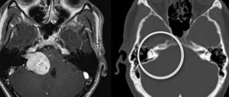
Atherosclerosis, critical stenosis of the internal carotid artery on CT angiogram (3D reconstruction)
Computed tomography is indicated for:
- regular headaches of unknown etiology;
- periodic loss of consciousness;
- dizziness;
- persistent increase in intracranial pressure;
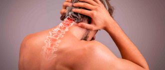
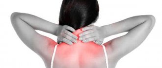
- pain and swelling in the neck;
- recurring attacks of nausea with vomiting;
- the appearance of tinnitus;
- pain in the eyes, blurred vision;
- numbness of arms, legs;
- convulsions;
- difficulties with coordination of movements;
- repeated loss of balance.
CT angiography of vessels is necessary for people who have suffered a stroke, heart attack, head injury, suffering from cerebral circulation disorders, vegetative-vascular dystonia, patients with a history of diagnoses such as brain contusion, encephalopathy, pituitary adenoma, epilepsy, Parkinson's disease, hydrocephalus.
CT angiogram of the vessels of the brain and neck - photo
This study is performed before brain surgery (it helps clarify the location of the main branches, which are important not to damage during the intervention) and to monitor the recovery process after surgical treatment.
A CT scan is prescribed if a magnetic resonance examination is not possible.
Treatment
Treatment of atherosclerosis begins with correction of diet and lifestyle changes.
All patients are recommended to lose weight, moderate physical activity, and quit smoking and alcohol. Often the cause of atherosclerosis is a nutritional factor, so diet plays a very important role. To reduce the concentration of cholesterol in the blood, it is important to avoid meat broths, fatty, salty, smoked foods, semi-finished products, spicy, spicy, fried foods, and limit the consumption of table salt.
Prohibited products include:
- sugar;
- confectionery, sweets;
- mayonnaise, tomato and other store-bought sauces;
- bakery products made from wheat flour;
- offal;
- sweet carbonated drinks;
- industrially produced juices and nectars;
- cocoa;
- canned meat and fish;
- dried fruits high in sugars;
- strong coffee and tea.
It is recommended to eat split meals five times a day, including a sufficient amount of fiber, protein (lean meats and fish, low-fat cottage cheese), fresh vegetables (except potatoes), fruits, and dairy products.
Drug therapy is aimed at:
- decreased cholesterol synthesis (statins, fibrates);
- decreased absorption of fats from food (cholesterol absorption inhibitors, bile acid sequestrants);
- prevention of thromboembolic complications (antiplatelet agents);
- relief of symptoms of atherosclerosis (painkillers, antispasmodics).
To determine indications for surgical treatment, the degree of disruption of blood flow in the vessel is determined.
An overlap of its lumen of less than 50% is considered hemodynamically insignificant; with stenosis of 50-70%, only drug therapy is usually carried out. Correction of arterial obstruction surgically must be performed in the third stage of atherosclerosis and narrowing of the lumen by more than 70%. There are two main types of surgical intervention:
- Balloon angioplasty. A balloon is inflated inside the affected vessel, while the atherosclerotic plaque is flattened and evenly distributed along the artery wall. At the end of the manipulation, the device is removed.
- Arterial stenting. During the operation, a thin lattice cylinder is inserted and opened in the area of narrowing. It also presses against the plaque, but remains in the lumen of the vessel, gradually growing into its endothelium.
Contraindications to CT angiography of the head and neck
Contraindications to computed tomography
Due to the use of iodine-based contrast agents, CT of the head and neck vessels has a number of limitations. The examination is contraindicated in the following situations:
- allergy to iodine and seafood, intolerance to the components of the amplifier: contrast can cause redness of the skin, rash, itching, difficulty breathing, bronchospasm, Quincke's edema, anaphylactic shock;
- exceeding the permissible value of creatinine in the blood: there is a high probability of kidney disease, in which the drug for improving the quality of CT photos of the brain and neck will not be removed from the body in time;
- treatment of diabetes with metformin: the drug reduces the rate of renal filtration and, together with iodine-containing drugs, can provoke a dangerous accumulation of lactic acid (lactic acidosis);
- hyperfunction of the thyroid gland: the introduction of contrast will increase the symptoms of the disease or provoke other complications (arrhythmia, etc.).
Due to the harmful effects of X-rays on the fetus, CT diagnostics of the vessels of the head and neck is not performed on pregnant women. According to strict indications, patients in the position can undergo a magnetic resonance examination. Unlike CT, MRI does not use ionizing radiation, therefore it is safer and is indicated even for children. But this diagnostic method is not suitable for those who have fixed metal dentures, braces, ear implants, etc.
The design features of the devices do not allow the procedure to be carried out if the person’s body weight is more than 150 kg or the chest (abdomen) circumference exceeds 150 cm. In this case, preference should be given to another research method, for example, magnetic resonance imaging using an open-type scanning device.
3D CT angiography of cerebral vessels
Congenital vasoconstriction
Narrowing of the neck vessels can be a congenital pathology, which is rarely isolated and usually develops against the background of other diseases, for example, vascular hypoplasia, abnormal development of the cervical vertebrae, vertebral arthrosis. Of great importance in the prevention of congenital defects of the spine is the intake of folic acid (vitamin B9) during the period of preparation for pregnancy and in the first trimester, when the formation of all vital organs of the fetus, including the neural tube, occurs.
Folic acid during pregnancy
Not only deficiencies of various vitamins, but also other factors (from the mother’s side), for example:
- infectious diseases of the mother, leading to infection of the amniotic fluid and amniotic membrane;
- intoxication of the fetus with decay products of ethyl alcohol, nicotine, ammonia and other toxic substances (if the mother has bad habits or the woman lives in environmentally unfavorable areas);
- injuries received during pregnancy.
Trauma during pregnancy
Deformation of the cervical vertebrae and the resulting narrowing of the neck vessels in a newborn may be the result of a protracted or difficult labor, especially if obstetric forceps or a vacuum aspirator were used to remove the baby.