What are the features of the symptoms of this disease?
In the presence of an ischemic stroke in the territory of the right middle cerebral artery, it is possible to identify symptoms of lesions of the midbrain and cerebral hemisphere, depending on the location and conditions of collateral blood supply in the clinical pictures. Quite often you can find a combination of damage to the thalamus and the cerebral hemisphere or isolated infarctions of the thalamus. It should be noted that in most cases, the symptoms of the disease in patients can be combined. The most common symptoms include visual damage, neuropsychological damage, and hemiparesis.
Diagnosis of stroke
The diagnosis is based on a thorough study of the medical history, identification of risk factors and analysis of clinical data, namely neurological symptoms. The clinical picture of strokes is varied and is largely determined by the vascular system in which the cerebral catastrophe occurred and its nature (ischemia or hemorrhage).
Early diagnosis of stroke (history and examination) involves answering the following questions:
— Does the patient have acute damage to the brain or spinal cord or their membranes?
— Is this lesion caused by a stroke?
ACVA is diagnosed with the sudden (minutes, less often hours) appearance of focal and/or general cerebral and meningeal neurological symptoms in a patient with a general vascular disease and in the absence of other causes, namely: traumatic brain or spinal injury; intoxication (alcohol, drugs, medications); hypoglycemia; infection; renal failure; liver failure.
Focal neurological symptoms are manifested by the occurrence of the following disorders:
– motor: mono-, hemi-, paraparesis – weakness in one or two limbs; paresis of the cranial nerves – facial asymmetry, hyperkinesis, etc.
– speech: sensory, motor aphasia – impaired understanding and pronunciation of words, dysarthria – unclear pronunciation of words, “porridge in the mouth”;
– sensitive: hypalgesia, thermoanesthesia, violation of deep, complex types of sensitivity, etc.;
– coordinators: vestibular, cerebellar ataxia, astasia, abasia – dizziness, unsteadiness when walking, falling to any side.
– visual: scotomas, quadrant and hemianopsia, amaurosis, photopsia – decreased or absent vision, loss of visual fields;
– cortical functions: astereognosis, apraxia – violation of the order of actions;
– memory: fixation amnesia, disorientation in time, etc.
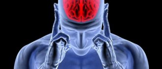
General cerebral symptoms: decreased level of wakefulness from subjective feelings of “vagueness,” “fogginess” in the head and mild stupor to deep coma; headache and pain along the spinal roots, nausea, vomiting.
Meningeal symptoms (can appear simultaneously with general cerebral and/or focal neurological symptoms, but more often appear somewhat delayed after the clinical debut of a stroke; with subarachnoid hemorrhages it can be the only clinical syndrome): tension of the posterior cervical muscles, positive Kernig, Brudzinsky signs (upper, middle, lower ), Bekhtereva, etc.
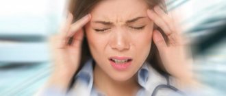
The most common first symptoms of a stroke are: sudden numbness or weakness in the arm and/or leg; sudden difficulty speaking and/or understanding it; sudden loss of balance, lack of coordination, dizziness; sudden loss of consciousness; severe headache for no apparent reason or after severe stress, physical overexertion sudden numbness of the lip or half of the face, often with “distortion” of the face.
Urgently call an ambulance by phone “03” if you see any of these symptoms in your friends or relatives or feel any of these symptoms yourself! A direct relationship between the outcome of a stroke and the time of initiation of its treatment has been proven. Every minute counts!
If a stroke is suspected, the patient must be hospitalized in a specialized neurological department for the treatment of patients with stroke.
What are the features of diagnosing ischemic stroke in the territory of the right middle cerebral artery?
It is worth noting that often computed tomography does not allow identifying any ischemic changes in the brain parenchyma for a certain moment after the onset of strokes, precisely the time that is very important as the beginning of the treatment of this type of disease.
Thanks to the use of magnetic resonance imaging, it becomes possible to more accurately determine the presence and nature of any ischemic changes in the main brain during strokes. After obtaining data from magnetic resonance imaging, it becomes possible to identify early ischemic changes. Today it has become possible to combine different modes, which makes it possible to determine more severe, subacute and congenital ischemic changes in the brain parenchyma.
Main services of Dr. Zavalishin’s clinic:
- consultation with a neurosurgeon
- treatment of spinal hernia
- brain surgery
- spine surgery
Differential diagnosis.
It should be remembered that in conditions such as hypoglycemia, uremia, liver failure, the sudden appearance of focal neurological disorders is possible. In case of infectious diseases, purulent diseases of the paranasal sinuses, the rapid development of focal/meningeal disorders is also possible, in this case indicating the addition of meningoencephalitis. One should not forget about traumatic brain injuries as the cause of loss of consciousness and the appearance of focal neurological disorders, especially in cases where the medical history is unknown and verbal contact with the patient is impossible; a careful examination and palpation of the soft tissues of the head, examination of the external auditory organs helps in the differential diagnosis and nasal passages for cerebrospinal fluid and hematorrhea; with subdural hematomas, anisocoria is detected in more than 90% of cases. With exogenous and endogenous intoxications (alcohol, ketoacidosis, etc.), leading to deep depression of consciousness, difficulties in differential diagnosis may also arise, since rare and shallow breathing does not allow the smell of alcohol, acetone, etc. to be detected in the exhaled air. Sudden appearance of focal neurological symptoms it is also possible with brain tumors - a “stroke-like” course of the disease or in the post-attack period in patients suffering from epileptic seizures (Todd’s palsy). In this condition, the resulting focal symptoms completely regress within 24 hours, and their appearance is caused not by “stroke” changes, but by local cerebral edema. In patients suffering from migraines, a sudden development of focal neurological symptoms (ophthalmic, oculomotor, sensory, motor, speech disorders, etc.) is also possible, the appearance of which may precede an attack of headache and disappear with the development of the migraine attack itself.
Diagnosis of the nature of the stroke
There are no characteristic clinical signs for hemorrhagic and ischemic strokes.
Clarification of the nature of the stroke is possible only after neuroimaging (computed tomography or magnetic resonance imaging).
Treatment of strokes.
Stroke therapy is divided into basic and specific.
Basic therapy for stroke is carried out regardless of the nature of stroke and begins from the first minutes of the patient’s admission to a specialized hospital.
— Normalization of vital functions - breathing and cardiovascular activity. If consciousness is not disturbed or depressed no deeper than stupor (the patient opens his eyes if called), it is necessary to sanitize the upper respiratory tract and ensure free breathing (elimination of factors that impede breathing - tight clothing, uncomfortable head position, recessed tongue, dentures, etc. .). If consciousness is depressed deeper than stupor, oxygen inhalation is necessary, and if breathing is inadequate, mechanical ventilation is necessary.
— The fight against high or low blood pressure is carried out in stages. Stabilization of elevated blood pressure at a level exceeding 20 mm Hg. the level familiar to the patient, and in the absence of anamnestic data, not lower than 150–160/90 mm Hg. Art.
— Combating cerebral edema (intracranial hypertension). Cerebral edema is defined as excess accumulation of fluid in the brain tissue, resulting in swelling and increased volume of the brain. But the bone walls of the skull are not extensible! Intracranial pressure increases sharply, which causes displacement of brain structures, and sometimes their compression. One of the main methods of treating cerebral edema is the administration of special medications - osmotic diuretics. They create high osmotic pressure in the blood, which forces fluid from the brain tissue along a pressure gradient into the blood, the flow of which carries it away from the cranial cavity.
— Maintaining water and electrolyte balance — the optimal ratio of fluid and electrolytes (potassium, sodium) in the body.
— Maintaining normal blood glucose levels (glycemia), since low (hypoglycemia) and high (hyperglycemia) blood glucose levels are equally dangerous for the brain, so it is necessary to maintain normoglycemia.
— Maintaining normal body temperature, since temperatures above 38*C aggravate brain damage, therefore temperature reduction is carried out both with medication and with the help of external cooling (water-alcohol rubdowns, ice packs). But be sure to find out the cause of the fever, since this may be a manifestation of an infection that requires completely different treatment.
— The fight against deep vein thrombosis in bedridden patients (the risk of pulmonary embolism and death of the patient) is carried out by elastic bandaging of the lower extremities, the use of pneumatic massagers, as well as the use of small doses of anticoagulants (heparin, fraxiparin), which reduce the tendency to thrombus formation.
— Relief of psychomotor agitation, epileptic seizures, nausea and vomiting, pain syndrome is carried out by special groups of drugs.
- Prevention of bedsores is mandatory - treating the patient’s skin, turning him over in bed, positioning the limbs with the help of special pillows. Rehabilitation of the patient begins as early as possible. Early rehabilitation – on days 2-3 when the patient’s condition is stable – significantly improves the outcome of stroke. This includes passive movements in the limbs for the prevention of contractures, verticalization of the patient using a special table - a verticalizer, in a stable condition - mobilization of the patient: transfer to a chair several times a day, physical therapy, physical therapy, for speech and swallowing disorders - speech therapy correction.
— The patient’s nutrition begins from 1-2 days. If swallowing is impaired (swallowing function is assessed immediately upon hospitalization and during treatment), a nasogastric tube is installed and nutritional mixtures are used for nutrition. If the patient is conscious and can swallow, then nutrition is carried out in the usual way.
Special methods for treating stroke.
Without an accurate determination of the nature of the stroke, it is impossible to carry out special treatment! The decision to use a particular drug or method can only be made by the patient’s attending physician. Treatment for a stroke will depend on the type of stroke.
1. In ischemic stroke, the main goals will be to restore cerebral blood flow and protect nerve cells from further damage. Thrombolytics (alteplase) are medications that dissolve blood clots. They are very effectively used in the treatment of myocardial infarction. By dissolving the clot and restoring blood flow, these drugs can prevent the development of damage to nerve cells and, therefore, cerebral infarction or significantly reduce its size. However, the use of such promising drugs is limited by the development of complications, the most serious of which is the “translation” of an ischemic stroke into a hemorrhagic one, i.e. secondary hemorrhage develops in the damaged brain tissue. Therefore, these drugs are indicated only for a strictly defined group of patients and can be used only in the earliest stages of stroke development (up to 4 hours from the onset). Anticoagulant drugs (warfarin) help stop the growth of existing blood clots and prevent the formation of new ones. This results in a decrease in blood clotting. This can lead to the development of internal bleeding; the risk is especially high in patients with gastric or duodenal ulcers and blood diseases. Another group of drugs that change the rheological properties of blood are called antiplatelet agents (aspirin). These drugs inhibit platelet sticking (aggregation). Antiplatelet agents are among the standard treatments for stroke caused by cerebral atherosclerosis and various blood diseases, which are based on its tendency to form blood clots due to platelet aggregation. These drugs are often used to prevent the development of both first and recurrent strokes. Thrombolytics, anticoagulants, and antiplatelet agents help restore and improve cerebral blood flow.
Unfortunately, without receiving the necessary oxygen and nutrients, brain cells begin to die. At the same time, a whole cascade of biochemical reactions is launched in them. Protective medications help stop this mechanism. They are called neuroprotectors, or cytoprotectors (from the Latin cytos - “cell”). Currently, serious clinical trials of drugs from this group are being conducted. It will be shown exactly how much the cerebral infarction is reduced when using these drugs. In addition, neuroprotectors help the cells surrounding the dead survive this “stress.” They increase their activity and even help some “volunteers” who were not previously involved in providing movement and/or speech, reorganize and, to a certain extent, take on the responsibilities of the dead.
Surgical methods for treating ischemic stroke include carotid endarterectomy. This operation is the method of choice if the cause of the stroke is severe stenosis or blockage of the carotid artery by an atherosclerotic plaque. It is also used to prevent the occurrence of both first and recurrent strokes. However, there are clearly defined indications and contraindications for this operation.
2. In hemorrhagic stroke, the main goals are to control blood pressure and stop bleeding. Lowering blood pressure helps reduce blood pressure on the walls of blood vessels, and bleeding stops faster. Blood pressure correction is carried out with antihypertensive (pressure-lowering) drugs that the patient takes orally (tablets and capsules) and which are administered intravenously to the patient. It all depends on the general condition of the patient, his blood pressure level and its fluctuations. It would seem that it is possible to perform an operation and “sew up” the ruptured vessel. However, this is not always possible and should be done. First, the bleeding may stop on its own if your blood pressure is controlled. Secondly, unfortunately, there are inoperable cases when the hemorrhage is very deep in the depths of the brain and the surgeon’s path to it is not easy. A neurosurgical operation is performed when it is possible to remove a sufficiently large hematoma, more than forty cubic centimeters, which compresses adjacent brain structures. This intervention can be carried out in two ways, open and closed. In the first case, neurosurgeons open the patient’s skull (perform trepanation) - an open intervention, and in the second case, using a special technique, they insert a cannula through a small hole into the very center of the hemorrhage and remove the spilled blood. The second option is called closed, or stereotactic, intervention. If the hemorrhage occurs from a rupture of an aneurysm, most often a congenital saccular protrusion on the wall of a vessel, then it is “sutured” with special metal clips - the aneurysm is clipped.
Life goes on after a stroke. But even after a relatively mild stroke, there is no need to succumb to “rosy” optimism. Any stroke is a serious warning about trouble in the body. Some advice for a patient who has suffered a stroke: Start recovery measures as quickly as possible: as soon as the general well-being of the patient and his state of consciousness allow it (for more details, see the section on the rehabilitation of a patient with a stroke). The patient’s relatives and friends should actively help him in conducting additional classes (in relation to the main ones conducted by the methodologist of therapeutic gymnastics and speech therapist-aphasiologist) in therapeutic gymnastics, in learning to walk and self-care, in restoring speech, reading and writing. The care of loved ones and a favorable psychological climate in the family should at the same time exclude “overprotection.” The patient must be given maximum independence, he must be encouraged to have an active social life. Return to active life, and where possible, to work, can occur not only with complete restoration of impaired functions, but also against the background of remaining partial motor and speech defects. And it is necessary to take measures to prevent a recurrent stroke (carefully read the guidelines on stroke prevention).
What is the procedure for treating ischemic stroke in the territory of the right middle cerebral artery?
To begin with, it is worth noting that the healing process is quite long and requires patience on the part of patients. At the very beginning of the healing process, you should normalize your lifestyle and attend rehabilitation events. The motor rehabilitation process includes strength and dexterity in each limb, self-care skills, all of which can be fully or partially rehabilitated. The speech rehabilitation process includes every session with specialists, in particular with speech therapists and neuropsychologists, every exercise necessary to restore common reading or counting disorders. As for the psychological and social healing processes, a healthy climate should be created in families, participation in any cultural event within social circles.
Quite often, specialists in this field of activity prescribe their patients to use various types of antidepressants, which are selected individually for each patient. Great attention should be paid to this, because using your own assumptions about taking antidepressants can only lead to various complications and side effects that can provoke undesirable consequences. That is why only the attending physician can prescribe the period of taking the drugs and the immediate dosage. The use of antiaggregates can reduce the risk of stroke recurrence, and in cases of no therapy, the disease may also return.
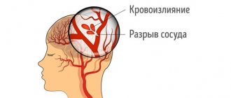
Stroke
Stroke
— acute cerebrovascular accident (ACVA), characterized by the sudden (within a few minutes, hours) appearance of focal and/or cerebral neurological symptoms, which persist for more than 24 hours or lead to the death of the patient in a shorter period of time due to cerebrovascular pathology.
Stroke includes cerebral infarction, cerebral hemorrhage and subarachnoid hemorrhage, which have etiopathogenetic and clinical differences.
Taking into account the time of regression of neurological deficit, transient cerebrovascular accidents (neurological deficit regresses within 24 hours, unlike the stroke itself) and minor stroke (neurological deficit regresses within three weeks after the onset of the disease) are especially distinguished. Vascular diseases of the brain occupy second place in the structure of mortality from diseases of the circulatory system after coronary heart disease.
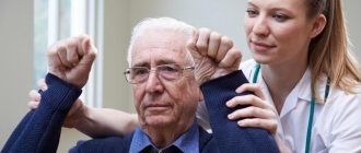
It is possible to recognize a stroke on the spot, immediately; For this, three main techniques for recognizing stroke symptoms, the so-called “USP” are used. To do this, ask the victim:
- U - smile. With a stroke, the smile may be crooked, the corner of the lips on one side may be directed downward rather than upward.
- Z - speak. Say a simple sentence, for example: “The sun is shining outside the window.” With a stroke, pronunciation is often (but not always!) impaired.
- P - raise both hands. If your arms don't rise at the same rate, this could be a sign of a stroke. Additional diagnostic methods:
- Ask the victim to stick out his tongue. If the tongue is curved or irregular in shape and falls to one side or the other, then this is also a sign of a stroke.
- Ask the victim to stretch his arms forward, palms up, and close his eyes. If one of them begins to involuntarily “move” sideways and downward, this is a sign of a stroke.
If the victim finds it difficult to complete any of these tasks, you must immediately call an ambulance and describe the symptoms to the doctors who arrived at the scene. Even if the symptoms have stopped (transient cerebrovascular accident), there should be one tactic - emergency hospitalization; old age and coma are not contraindications to hospitalization.
There is another mnemonic rule for diagnosing a stroke: U.D.A.R.
.:
- U - Smile After a stroke, the smile becomes crooked and asymmetrical;
- D - Movement Raise both arms and both legs up at the same time - one of the paired limbs will rise slower and lower;
- A - Articulation Say the word “articulation” or several phrases - after a stroke, diction is disrupted, speech sounds inhibited or simply strange;
- R - Decision If you find violations in at least one of the points (compared to the normal state), it’s time to make a decision and call an ambulance. Tell the dispatcher what signs of a stroke (STROKE) you have found and a special resuscitation team will arrive quickly.
Types of stroke
There are three main types of stroke:
- Ischemic stroke,
- Intracerebral hemorrhage.
- Subarachnoid hemorrhage.
Intracerebral and (not in all classifications) non-traumatic intrathecal hemorrhages are classified as hemorrhagic stroke. According to multicenter studies, the ratio of ischemic and hemorrhagic strokes averages 4:1-5:1 (80-85% and 15-20%).
Ischemic stroke.
Ischemic stroke, or cerebral infarction.
Most often it occurs in patients over 60 years of age with a history of myocardial infarction, rheumatic heart disease, cardiac arrhythmia and conduction disorders, and diabetes mellitus. Violations of the rheological properties of blood and pathology of the main arteries play a major role in the development of ischemic stroke. The development of the disease at night without loss of consciousness is typical.
Etiopathogenesis.
Ischemic stroke most often occurs when the arteries supplying the brain become narrowed or blocked. Without receiving the oxygen and nutrients they need, brain cells die. Ischemic stroke is divided into atherothrombotic, cardioembolic, hemodynamic, lacunar and hemorheological microocclusion type stroke.
- Atherothrombotic stroke, as a rule, occurs against the background of atherosclerosis of cerebral arteries of large or medium caliber. Atherosclerotic plaque narrows the lumen of the vessel and promotes thrombus formation. Possible arterio-arterial embolism. This type of stroke develops in stages, with an increase in symptoms over several hours or days, and often debuts during sleep. Often, atherothrombotic stroke is preceded by transient ischemic attacks. The size of the focus of ischemic damage varies.
- Cardioembolic stroke occurs when a cerebral artery is completely or partially blocked by an embolus. The most common causes of stroke are cardiogenic embolism due to valvular heart disease, recurrent rheumatic and bacterial endocarditis, and other heart lesions that are accompanied by the formation of parietal thrombi in its cavities. Often, embolic stroke develops as a result of paroxysm of atrial fibrillation. The onset of cardioembolic stroke is usually sudden, while the patient is awake. At the onset of the disease, the neurological deficit is most pronounced. More often, a stroke is localized in the area of the blood supply of the middle cerebral artery, the size of the focus of ischemic damage is medium or large, and a hemorrhagic component is characteristic. There may be a history of thromboembolism in other organs.
- Hemodynamic stroke is caused by hemodynamic factors - a decrease in blood pressure (physiological, for example during sleep; orthostatic, iatrogenic arterial hypotension, hypovolemia) or a drop in cardiac output (due to myocardial ischemia, severe bradycardia, etc.). The onset of a hemodynamic stroke can be sudden or gradual, while the patient is at rest or active. The sizes of infarctions vary; localization is usually in the area of adjacent blood supply (cortical, periventricular, etc.). Hemodynamic strokes occur against the background of pathology of extra- and/or intracranial arteries (atherosclerosis, septal artery stenosis, abnormalities of the cerebral vascular system).
- Lacunar stroke is caused by damage to small perforating arteries. As a rule, it occurs against a background of high blood pressure, gradually, over several hours. Lacunar strokes are localized in subcortical structures (subcortical nuclei, internal capsule, white matter of the semioval center, base of the bridge), the size of the lesions does not exceed 1.5 cm. There are no general cerebral and meningeal symptoms, there are characteristic focal symptoms (purely motor or purely sensory lacunar syndrome, atactic hemiparesis, dysarthria or monoparesis).
- Stroke of the hemorheological microocclusion type occurs in the absence of any vascular or hematological disease of established etiology. The cause of stroke is pronounced hemorheological changes, disturbances in the hemostasis and fibrinolysis systems. Characterized by scant neurological symptoms in combination with significant hemorheological disorders. Hemorrhagic stroke. In the scientific literature, the terms “hemorrhagic stroke” and “non-traumatic intracerebral hemorrhage” are either used as synonyms, or hemorrhagic strokes, along with intracerebral strokes, also include non-traumatic subarachnoid hemorrhage.
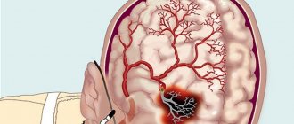
- Intracerebral hemorrhage is the most common type of hemorrhagic stroke, most often occurring between the ages of 45 and 60 years. Such patients have a history of hypertension, cerebral atherosclerosis or a combination of these diseases, arterial symptomatic hypertension, blood disease, etc. Precursors of the disease (feeling hot, increased headache, blurred vision) are rare. Typically, a stroke develops suddenly, during the daytime, against the background of emotional or physical stress. Etiopathogenesis. The cause of cerebral hemorrhage is most often hypertension (80-85% of cases). Less commonly, hemorrhages are caused by atherosclerosis, blood diseases, inflammatory changes in cerebral vessels, intoxication, vitamin deficiencies and other reasons. Cerebral hemorrhage can occur through diapedesis or as a result of rupture of a vessel. In both cases, the release of blood beyond the vascular bed is based on functional-dynamic angiodystonic disorders of the general and especially regional cerebral circulation. The main pathogenetic factor of hemorrhage is arterial hypertension and hypertensive crises, in which spasms or paralysis of cerebral arteries and arterioles occur. Metabolic disorders that occur in the focus of ischemia contribute to the disorganization of the walls of blood vessels, which under these conditions become permeable to plasma and red blood cells. This is how hemorrhage occurs through diapedesis. The simultaneous development of spasm of many vascular branches in combination with the penetration of blood into the medulla can lead to the formation of an extensive focus of hemorrhage, and sometimes multiple hemorrhagic foci. The basis of a hypertensive crisis may be a sharp expansion of the arteries with an increase in cerebral blood flow, caused by a breakdown in its self-regulation with high blood pressure. Under these conditions, the arteries lose their ability to narrow and passively expand. Under increased pressure, blood fills not only the arteries, but also the capillaries and veins. At the same time, vascular permeability increases, which leads to diapedesis of blood plasma and erythrocytes. In the mechanism of occurrence of diapedetic hemorrhage, a certain importance is attached to the disruption of the relationship between the coagulation and anticoagulation systems of the blood. Functional-dynamic disturbances of vascular tone also play a role in the pathogenesis of vascular rupture. Paralysis of the walls of small cerebral vessels leads to an acute increase in the permeability of the vascular walls and plasmorrhagia.
- Subarachnoid hemorrhage - (hemorrhage into the subarachnoid space). Most often, hemorrhage occurs between the ages of 30 and 60 years. Risk factors for the development of subarachnoid hemorrhage include smoking, chronic alcoholism and single consumption of alcohol in large quantities, arterial hypertension, and excess body weight.
Etiopathogenesis.
It can occur spontaneously, usually due to rupture of an arterial aneurysm (according to various sources, from 50% to 85% of cases) or as a result of traumatic brain injury. Hemorrhages are also possible due to other pathological changes (arteriovenous malformations, diseases of the spinal cord vessels, hemorrhage into the tumor). In addition, causes of SAH include cocaine addiction, sickle cell anemia (usually in children); less often - taking anticoagulants, blood coagulation disorders and pituitary stroke. The location of subarachnoid hemorrhage depends on the location of the vessel rupture. Most often it occurs when the vessels of the arterial circle of the cerebrum on the lower surface of the brain rupture. An accumulation of blood is detected on the basal surface of the cerebral peduncles, pons, medulla oblongata, and temporal lobes. Less commonly, the lesion is localized on the superolateral surface of the brain; the most intense hemorrhages in these cases can be traced along the large grooves.
Clinical picture.
Symptoms: Stroke can manifest itself with general cerebral and focal neurological symptoms. General cerebral symptoms of stroke vary. This symptom may occur in the form of impaired consciousness, stupor, drowsiness, or, conversely, agitation; a short-term loss of consciousness may also occur for several minutes. A severe headache may be accompanied by nausea or vomiting. Sometimes dizziness occurs. A person may feel a loss of orientation in time and space. Possible vegetative symptoms: feeling of heat, sweating, palpitations, dry mouth.
Against the background of general cerebral symptoms of stroke, focal symptoms of brain damage appear. The clinical picture is determined by which part of the brain is affected due to damage to the blood vessel supplying it. If a part of the brain provides the function of movement, then weakness in the arm or leg develops, including paralysis. Loss of strength in the limbs may be accompanied by a decrease in sensitivity in them, impaired speech, and vision. These focal stroke symptoms are mainly associated with damage to the area of the brain supplied by the carotid artery. Muscle weakness (hemiparesis), speech and pronunciation disorders, decreased vision in one eye and pulsation of the carotid artery in the neck on the affected side are characteristic. Sometimes there is unsteadiness of gait, loss of balance, uncontrollable vomiting, dizziness, especially in cases where the blood vessels supplying the areas of the brain responsible for coordinating movements and a sense of body position in space are affected. “Spotty ischemia” occurs in the cerebellum, occipital lobes and deep structures and brain stem. Attacks of dizziness in any direction are observed when objects rotate around a person. Against this background, there may be visual and oculomotor disturbances (strabismus, double vision, decreased visual fields), unsteadiness and instability, deterioration in speech, movements and sensitivity.
Risk factors.
Risk factors are various clinical, biochemical, behavioral and other characteristics that indicate an increased likelihood of developing a certain disease. All areas of preventive work are focused on controlling risk factors and their correction both in specific people and in the population as a whole.
- Age.
- Arterial hypertension.
- Heart diseases.
- TIAs (transient ischemic attacks) are a significant predictor of the development of both cerebral infarction and myocardial infarction.
- Diabetes.
- Smoking.
- Asymptomatic stenosis of the carotid arteries.
Many people in the population have several risk factors simultaneously, each of which can be moderately expressed.
Diagnostics.
Computed tomography (CT) and magnetic resonance imaging (MRI) are the most important diagnostic tests for stroke. In most cases, CT allows one to clearly differentiate “fresh” cerebral hemorrhage from other types of strokes; MRI is preferable for identifying areas of ischemia, assessing the extent of ischemic damage and penumbra (this is especially important in the first 12-24 hours of the disease, when an ischemic stroke may not be detected by CT). be visualized). These studies can also detect primary and metastatic tumors, brain abscesses and subdural hematomas. If there is a stiff neck but no papilledema, a lumbar puncture will provide a rapid diagnosis of cerebral hemorrhage in most cases, although there is still a slight risk of brain herniation syndrome. In cases where embolism is suspected, lumbar puncture is necessary if anticoagulants are to be used. Lumbar puncture is also important for the diagnosis of multiple sclerosis and, in addition, can be of diagnostic value in neurovascular syphilis and brain abscess. If CT or MRI is not available, echoencephalography and lumbar puncture should be performed.
First aid for stroke.
In the case of a stroke, the most important thing is to get the person to hospital as quickly as possible, preferably within the first hour after symptoms are noticed. It should be borne in mind that not all hospitals, but only a number of specialized centers are equipped to provide proper modern stroke care. Therefore, attempts to independently transport a patient to the nearest hospital during a stroke are often ineffective. Before the ambulance arrives, it is important not to allow the patient to eat or drink, since the swallowing organs may be paralyzed, and then food entering the respiratory tract can cause suffocation. At the first signs of vomiting, the patient's head is turned to the side so that the vomit does not enter the respiratory tract. It is better to lay the patient down with pillows under his head and shoulders, so that the neck and head form a single line, and this line makes an angle of about 30° to the horizontal. The patient should avoid sudden and intense movements. The patient is unbuttoned from tight, obstructive clothing, his tie is loosened, and his comfort is taken care of.
In case of loss of consciousness with absent or agonal breathing, cardiopulmonary resuscitation is started immediately. Its use greatly increases the patient's chances of survival. The use of portable defibrillators further increases survivability: when in a public place (cafe, airport, etc.), first aid providers need to ask the staff if they have a defibrillator or nearby. Determining the absence of a pulse is no longer a necessary condition for starting resuscitation; loss of consciousness and absence of rhythmic breathing are sufficient.
Treatment:
Treatment of stroke includes a set of emergency measures and a long recovery period (rehabilitation), carried out in stages.
Urgent Care.
At the prehospital stage of medical care, the patient’s hemodynamic parameters should be assessed; if an increase in blood pressure is observed, measures should be taken to normalize it.
Resuscitation measures.
Resuscitation measures should be aimed at maintaining adequate hemodynamics and oxygenation.
Pharmacotherapy.
Drugs are prescribed according to treatment standards and according to the decision of the attending physician. Acids in drugs or food should be excluded in cases of hemorrhage as they help weaken blood clotting and, conversely, are recommended for ischemic strokes as they help resolve thrombosis.
Features of patient care.
Stroke is often accompanied by pneumonia and bedsores, which require constant care, turning from side to side, changing wet underwear, feeding, bowel cleansing, and percussion chest massage.
Post-stroke rehabilitation.
Treatment of stroke includes a course of vascular therapy, the use of drugs that improve brain metabolism, oxygen therapy, restorative treatment or rehabilitation (physical therapy, physiotherapy, massage). It is also recommended to do breathing exercises for the lungs after being discharged from the hospital, since after 14-17 days of “lying” in the lungs, “congestive pneumonia” can form. That is, take deep breaths and exhales. Inflate balloons 5-7 times a day. In the world practice of rehabilitation treatment after a stroke, the leading place is occupied by an interdisciplinary approach, based on which several specialists are involved in the treatment (therapy) process: a physical therapy doctor, a rehabilitation specialist, a speech therapist.
The human brain has a certain natural ability to recover, thanks to the creation of new connections between healthy neurons and the formation of new information circuits. This property of the brain is called neuroplasticity and can be stimulated during the rehabilitation process. One of the key factors in the effectiveness of any rehabilitation program is the regular implementation of a carefully organized, individually selected set of exercises - that is, the general principle of teaching a person a new skill.
For information about making an appointment with specialists, please contact us by phone: 8;, or by email. The email address is being protected from spambots. Javascript must be enabled in your browser to view the address. / The email address is protected from spambots. Javascript must be enabled in your browser to view the address.