The structural unit of the nervous system is the nerve cell, or neuron. Even if one neuron is isolated from the rest of the nervous system, it retains the ability to perceive information, analyze it, and respond adequately. The nerve cell that is involved in the perception of information is called an afferent neuron, or sensory neuron. It is one of the main links of the reflex arc. More details about these structures are provided later in the article.
Neuron structure
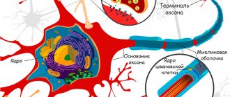
Fundamentally, the structure of the afferent neuron is no different from other cells of the nervous system. It has two main components: the body and processes. The shoots, in turn, are of two types:
- dendrites - have a relatively short length;
- an axon is a long extension of a neuron, always one per cell.
The function of dendrites is to transport nerve impulses to the nerve cell body. Due to this, information from the environment enters the nervous system. Dendrites also make it possible for neurons to communicate with each other.
A long process - an axon - carries information from the cell body to other parts of the body. In this way, information spreads beyond the nervous system.
The body of the afferent neuron consists of several parts:
- a shell consisting of phospholipids and proteins;
- nucleus with genetic material;
- the Golgi complex, which performs the function of packaging carbohydrates;
- endoplasmic reticulum, where protein synthesis occurs;
- mitochondria, in which ATP energy is generated;
- lysosomes, which perform the functions of “swallowing” foreign particles.
All these elements work together and are in constant interaction.
How do they work together and how are they different?
Afferent neurons typically have two axons that carry electrochemical signals to the spinal column or brain. Once there, the signal travels through a network of interneurons and through the efferent neuron. Afferent-efferent pairs of neurons that pass through the spine control reflexes (such as the knee-jerk reaction).
Finished works on a similar topic
Course work Afferent and efferent nerve conductors and their role in psychology 440 ₽ Abstract Afferent and efferent nerve conductors and their role in psychology 220 ₽ Test work Afferent and efferent nerve conductors and their role in psychology 250 ₽
Receive completed work or specialist advice on your educational project Find out the cost
Afferent neurons are designed to respond to various stimuli. For example, an afferent neuron designed to respond to heat detects excess heat and sends an impulse through the central nervous system. The efferent neuron then causes the muscles to contract to move the body away from the heat. The skin has sensory receptors for heat, cold, pleasure, pain and pressure.
Afferent neurons have round and smooth cell bodies, while efferent neurons have satellite bodies. Afferent neurons are found in the peripheral nervous system, and efferent neurons are located in the central nervous system. Axons in afferent neurons move from the ganglia (clusters of nerve cells that contain afferent and efferent neurons) to the spinal cord. The long axon is actually connected to the efferent neuron.
Afferent neurons have a single long myelinated dendrite, whereas efferent neurons have shorter dendrites. The dendrite in an afferent neuron is what is responsible for transmitting nerve impulses from the receptors to the cell body, while in an efferent neuron the impulses travel through the dendrite and exit through the neuromuscular junction that is formed between the effectors and the axon.
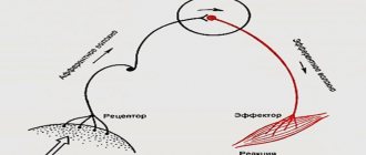
Concept and structure of the reflex arc
A reflex is a universal response of the body to irritation. The unconditioned reflex is transmitted hereditarily and is the same for any person. Conditional is one that is acquired during life. Each person can have his own conditioned reflex.
Afferent and efferent neurons are the main components of the reflex arc.
At its core, a reflex arc is a path that overcomes a nerve impulse to a working organ or muscle. There are three main components of the arc:
- analyzer, or afferent, part;
- central, or contact, part;
- executive, or efferent, part.
According to modern concepts, there are not three, but five parts of the reflex arc. It begins with a receptor - the end of a sensitive neuron. In addition to the receptor, the arc includes the following elements:
- sensitive, or afferent, neuron;
- interneuron;
- motor, or efferent, neuron;
- the organ that responds to stimulation is the effector.
That is, for a reflex arc to form, there must be at least three nerve cells. An exception is the tendon reflexes, which contain only two cells - sensory and motor.
Analyzer part
An afferent neuron is a neuron that perceives information from the internal and external environment of the body using a receptor. Thus, the analyzer part consists of a receptor and a sensitive pathway.
There are several classifications of receptors. Depending on their location they are divided:
- on exteroceptors - those that are located in the skin and mucous membranes;
- proprioceptors - those located in muscle fibers, tendons and ligaments;
- interoreceptors - those that are located in the internal organs.
Depending on the type of stimulus that the receptor perceives, they are divided:
- to thermoreceptors - they react to changes in temperature, they are found on the skin and tongue;
- baroreceptors - react to pressure changes, located in the aortic arch, carotid sinus;
- chemoreceptors - react to changes in chemical composition, located in the stomach and intestines;
- photoreceptors - react to light, located in the retina;
- pain receptors - provide the perception of pain, found in the skin, peritoneum, capsule of internal parenchymal organs, periosteum;
- phonoreceptors - respond to sounds, located in the inner ear.
The sensitive pathway is the same afferent neuron with a body and processes. The axon of a nerve cell enters the central nervous system through the dorsal horns of the spinal cord, which are also called sensory. Directly in the spinal cord it transmits information to the interneuron.
Content
- Neurons Types of neurons
- Nerve fibers and nerves
- List of cranial nerves with dominant fiber designation
To get started, I recommend watching a short video that talks about different human tissues. But it is the nervous tissue that will interest us. In a more colorful and visual form, it will be easier for you to grasp the basics, and then you can expand your knowledge.
The main tissue from which the nervous system is formed is nervous tissue, which consists of cells and intercellular substance. Tissue is a collection of cells and intercellular substance that are similar in structure and functions.
Nervous tissue is of ectodermal origin. Nervous tissue differs from other types of tissue in that it lacks intercellular substance. The intercellular substance is a derivative of the glial cell and consists of fibers and amorphous substance.
The function of nervous tissue is to ensure the receipt, processing and storage of information from the external and internal environment, as well as the regulation and coordination of the activities of all parts of the body.
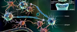
Nerve tissue consists of two types of cells: neurons and glial cells . Neurons play a major role in providing all functions of the central nervous system. Glial cells have an auxiliary role, performing supporting, protective, trophic functions, etc. On average, the number of glial cells exceeds the number of neurons in a ratio of 10:1, respectively.
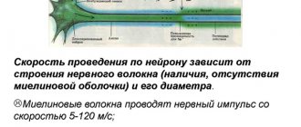
Each neuron has an expanded central part: a body - soma and processes - dendrites and axons . Impulses travel through dendrites to the nerve cell body, and through axons from the nerve cell body to other neurons or organs.
The shoots can be long or short. The long processes of neurons are called nerve fibers . Most dendrites (dendron - tree) are short, highly branched processes. The axon (axis - process) is often a long, slightly branched process.
Contact part
The contact part of the reflex arc is also called the nerve center. The number of interneurons may vary. It depends on the complexity of the information that needs to be processed. The interneuron is located within the central nervous system, namely the spinal cord.
The functions of the contact part are as follows:
- analysis of the information received;
- synthesis of information;
- making the final decision about what the answer should be.
Location of the sensory neuron
Afferent neurons are part of the peripheral nervous system. Their perceiving sections - receptors - are located in the skin, the walls of tubular organs or blood vessels, and the capsules of parenchymal organs.
Afferent neurons are pseudounipolar. That is, they have one process that extends from the body and then divides into two. Thus, one process approaches the body from the receptor, and then another extends from it to the center.
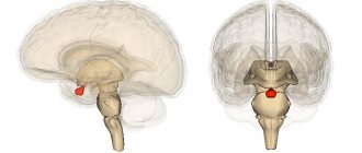
The body itself is located in the spinal ganglia, or nodes. These formations consist entirely of nerve cell bodies.
Concept and types of neurons
Definition 1
A neuron is an electrically excitable cell, a functional unit of the nervous system.
Each neuron has a cell body, dendrites, and an axon. Neurons are divided into three types:
- afferent neurons,
- efferent neurons
- interneurons.
Sensory information is transmitted from the periphery of the body to the main organ - the brain. Sensory information includes nerve impulses (that is, things people hear, touch, see, taste, and smell) that are transmitted from the senses. Afferent neurons are also called sensory neurons, and it is these specialized cells that transmit nerve impulses from the body directly to the central nervous system.
Are you an expert in this subject area? We invite you to become the author of the Directory Working Conditions
Physical stimuli, such as sound or light, activate afferent neurons, converting modalities into nerve impulses. They do this by using sensory receptors found in their cell membranes. The main cell bodies of afferent neurons are located near the brain and spinal cord, which together form the central nervous system.
Efferent neuron cells are located in the central nervous system and are called motor neurons. After receiving input from various neurons, including afferent neurons and interneurons, efferent neurons receive these signals from the central nervous system and transmit nerve impulses to the peripheral nervous system, muscles and glands to initiate a response to a stimulus.
Functions of the sensory neuron
The main function of the afferent neuron has already been noted above - the perception of information from the outside using a receptor. But what does this phrase mean?
The perception of any signal is based on the process of information analysis. Its essence lies in the detailed “decomposition” of the stimulus into individual components, careful study and identification of its individual properties. But only a superficial analysis of information occurs in the receptor. Therefore, one reflex arc is not capable of providing complex reactions. For example, one of the reflexes is extension of the knee to a blow to the tendon with a hammer.
A more subtle “decomposition” of the stimulus is carried out in the central nervous system. And this process is ensured most subtly and thoroughly by the cerebral cortex as the most historically new structure of the central nervous system.
Thus, the afferent neuron is an important part of the reflex arc, thanks to which we are able to perceive external irritation and quickly respond to it.
Efferent neurons
The motor centers of the cortex and spinal cord are characterized by excitation of interneurons due to feedback.
Interneurons can be excitatory or inhibitory.
Interneurons, their role in the formation of neural networks
Afferent neurons, their functions
Afferent neurons are neurons that perceive information.
As a rule, afferent neurons have a large branched network. This is typical for all levels of the central nervous system. In the posterior horns of the spinal cord, afferent neurons are small in size with a large number of dendritic processes, while in the anterior horns of the spinal cord, efferent neurons have a large body, coarser, less branching processes. These differences increase as the level of the central nervous system changes to the medulla oblongata, midbrain, diencephalon, and telencephalon. The greatest differences between afferent and efferent neurons are observed in the cerebral cortex.
Interneurons, or interneurons, process information received from afferent neurons and transmit it to other interneurons or efferent neurons.
Interneurons, as a rule, have axons whose terminals end on the neurons of their own center, primarily ensuring their integration.
Some interneurons receive activation from neurons in other centers and then distribute this information to neurons in their center. This ensures an increase in the influence of the signal due to its repetition in parallel paths and lengthens the time of storing information in the center. As a result, the center where the signal arrived increases the reliability of the influence on the executive structure.
Other interneurons receive activation from collaterals of efferent neurons in their own center and then transmit this information back to their own center, forming feedback connections. This is how reverberating networks are organized, allowing information to be stored in the nerve center for a long time.
Inhibitory interneurons are excited by direct signals going to their own center, or by signals coming from the same center, but via feedback connections. Direct excitation of inhibitory interneurons is characteristic of the intermediate centers of the afferent spinocerebral pathways.
Efferent neurons of the nervous system are neurons that transmit information from the nerve center to the executive organs or other centers of the nervous system. For example, the efferent neurons of the motor zone of the cerebral cortex - pyramidal cells, send impulses to the motor neurons of the anterior horns of the spinal cord, i.e. they are efferent for this part of the cerebral cortex. In turn, the motor neurons of the spinal cord are efferent to its anterior horns and send signals to the muscles. The main feature of efferent neurons is the presence of a long axon with a high speed of excitation.
Efferent neurons of different parts of the cerebral cortex connect these parts with each other via arcuate connections. Such connections provide intrahemispheric and interhemispheric relationships that form the functional state of the brain in the dynamics of learning, fatigue, pattern recognition, etc. All descending tracts of the spinal cord (pyramidal, rubrospinal, reticulospinal, etc.) are formed by the axons of efferent neurons of the corresponding parts of the central nervous system.
Neurons of the autonomic nervous system, for example, the nuclei of the vagus nerve, the lateral horns of the spinal cord, also belong to the efferent ones.
Neuroglia, or glia,
- a set of cellular elements of nervous tissue formed by specialized cells of various shapes. It was discovered by R. Virchow and he named it neuroglia, which means “nerve glue”. Neuroglial cells fill the spaces between neurons, making up 40% of the brain volume. Glial cells are 3-4 times smaller in size than nerve cells; their number in the central nervous system of mammals reaches 140 billion. With age in the human brain, the number of neurons decreases, and the number of glial cells increases.
There are several types of neuroglia, each of which is formed by cells of a certain type: astrocytes, oligodendrocytes, microgliocytes (see Fig. 5).
Astrocytes are multi-processed cells with oval-shaped nuclei. It is believed that astrocytes serve as a support for neurons, provide reparative processes in nerve trunks, isolate nerve fibers, and participate in the metabolism of neurons.
Astrocytes form bridges between the capillaries and the ependyma lining the cavities of the ventricles of the brain. It is believed that this ensures exchange between the blood and the cerebrospinal fluid of the ventricles of the brain, i.e., astrocytes perform a transport function. Apparently, they ensure the transport of substances from the blood to the neuron and back.
Oligodendrocytes are cells with a small number of processes. They are smaller in size than astrocytes. In the cerebral cortex, the number of oligodendrocytes increases from the upper layers to the lower. There are more oligodendrocytes in the subcortical structures and in the brain stem than in the cortex. Oligodendrocytes are involved in the myelination of axons (therefore there are more of them in the white matter of the brain), in the metabolism of neurons, as well as in the trophism of neurons.
Microglia are represented by the smallest multi-processed glial cells belonging to the wandering cells. The source of microglia is the mesoderm. Microglial cells are capable of phagocytosis.
Normal physiological processes in the nervous system largely depend on the degree of myelination of nerve cell fibers. In the central nervous system, myelination is provided by oligodendrocytes, and in the peripheral nervous system by lemmocytes (Schwann cells).
Glial cells do not have impulse activity, like nerve cells, but the membrane of glial cells has a charge that forms a membrane potential, which is very inert.
The membrane potential of neuroglia is 70-90 mV.
Glial cells are capable of transmitting excitation, the spread of which from one cell to another occurs with decrement.