Definition
Benign paroxysmal positional vertigo (BPPV) occurs in repeated episodes, often lasting less than one minute. Attacks are triggered by changes in head position: turning, throwing back, as well as changes in body position, including lying down, even in sleep. Between attacks, autonomic disturbances (nausea, rarely vomiting, fluctuations in blood pressure, sweating) and imbalance may persist, so patients may describe constant dizziness.
Over time, the severity of attacks usually decreases. The word “benign” means that the disease goes away on its own, without treatment, without causing lasting harm to the patient.
BPPV is the most common type of dizziness. Attacks most often develop in older women. However, the disease can occur at any age.
Attacks of BPPV, in most cases, are associated with separation, destruction or increase in size of the otoliths.
Otoliths (otoconia) are layered pebbles consisting primarily of calcium carbonate crystals, like mother of pearl or nacre. They are immersed in a jelly-like layer that envelops the hairs of sensitive cells on the surface of the macula (macula) of the spherical and eleptic sacs of the vestibular analyzer. The otoliths, the jelly-like layer and the hairs of sensory cells form the otolithic membrane.
The elliptic sac (uterus) connects to three semicircular tubules (SCC), located in three perpendicular planes: lateral, anterior and posterior. In their extensions at the junction with the uterus, there is also a sensitive area - the ampullar ridge, covered with a structure similar to the otolithic membrane - the cupula. Normally, the cupula separates the RCC and the utricle. It does not contain otoliths. The cupula provides the perception of angular accelerations of the head, responding to changes in pressure in the ampulla arising due to the inertia of the endolymph (the fluid that fills the RCC and the sacs of the vestibular analyzer).
Detached otoliths or fragments thereof may enter the ampullae of the RCC and irritate the cupular areas. This more common variant of BPPV is called canalithiasis.
Thanks to the balance between the formation and resorption of the layers that make up the otoliths, their renewal is ensured, as well as the resorption of detached otoliths. When the balance is disturbed, one of the otoliths becomes large (2-4 times larger than neighboring cells); the large mass leads to greater displacement compared to neighboring fixed otoliths, which is a source of irritation of the vestibular system. This variant of BPPV is called dome lithiasis; it is characterized by a longer course (several months) and lack of effect from vestibular maneuvers.
Asymmetrical signal input to the brain during unilateral stimulation of the vestibular apparatus disrupts the illusion of balance created by the interaction of the vestibular, visual and proprioceptive systems (receiving signals from muscles and ligaments, assessing the position of limb segments). There is a feeling of dizziness.
Sensitive cells of the vestibular analyzer send a signal of maximum intensity to the brain during the first second of stimulation, then the signal strength decreases exponentially, which underlies the short duration of BPPV symptoms.
The most common lesion is the posterior SCC (90%), less often the lateral one (8%), the remaining cases are caused by damage to the anterior SCC and combined damage to several tubules. Classic cases of BPPV due to posterior RCC involvement are idiopathic in 35% of cases, with previous traumatic brain injury (sometimes minor) and whiplash in the neck occurring in 15% of patients.
In other cases, BPPV is caused by other disorders: most often Meniere's disease (30%), vestibular neuronitis, surgical interventions on the organ of hearing, paranasal sinuses, herpetic lesions of the ear ganglion and circulatory disorders of the structures of the inner ear. Population studies have revealed a direct relationship between the likelihood of developing BPPV and age, female gender, migraine, giant cell arteritis, risk factors for cardiovascular complications - arterial hypertension and dyslipidemia, as well as a history of stroke, which confirms the importance of vascular causes in some cases.
Lindsay-Hemenway syndrome has been identified - acute dizziness, followed by the development of BPPV attacks and a decrease or complete disappearance of nystagmus in the caloric test due to circulatory disorders in the anterior vestibular artery system.
The diagnosis of BPPV is made based on the assessment of nystagmus during special maneuvers - techniques that cause angular accelerations of the patient's head.
Damage to the posterior semicircular tubule
The Dix-Hallpike test is the “gold standard” for diagnosing BPPV caused by posterior SCC pathology:
- The patient sits straight along the couch, with his head turned 45˚ towards the labyrinth that is being examined.
- The patient is placed in a lying position, while the head rotation is maintained, the head is thrown back at an angle of 30˚ relative to the axis of the body, and hangs over the edge of the couch.
- Watch eye movements. Nystagmus and dizziness occur with a delay of several seconds and last less than 1 minute. Nystagmus has a typical trajectory: first, a tonic phase occurs, during which the eyeball is moved upward, away from the underlying ear, a rotatory component is noted, then clonic eye movements toward the floor occur. the underlying ear.
- After the nystagmus stops, the patient is returned to a sitting position and the eye movements are observed again; the nystagmus may reappear, but in the opposite direction.
When the test is repeated with turning the head in the same direction, the intensity and duration of the nystagmus decrease each time.
The procedure is repeated with the head turned in the opposite direction.
The affected side is determined by which side positional nystagmus and dizziness occur.
Anatomy
The inner ear is represented by a labyrinth located in the temporal bone. The labyrinth consists of three semicircular canals (anterior, posterior, horizontal) and the cochlea. The semicircular canals are located in three mutually perpendicular planes: the horizontal (HPC) is located at an angle of 30° to the horizontal plane, and the anterior (APC) and posterior (PPC) are located at an angle of 45° to the sagittal plane. The hairs of the sensory cells of the otolith receptor form a network that is immersed in a jelly-like mass containing otoliths - crystals of phosphate and calcium carbonate, which together form the otolith membrane.
According to one theory of the occurrence of BPPV, an attack of dizziness occurs when the otoliths separate from the membrane and enter the lumen of one or more semicircular canals. During the displacement of otoliths in the lumen of the semicircular canal, under the influence of gravity when turning the head, movement of the endolymph occurs, which leads to excitation of nerve endings, the appearance of nystagmus (involuntary oscillatory movement of the eyes) and dizziness.
With BPPV, each of the semicircular canals can be affected, and in some cases several canals at once.
Most often (85-90% of cases) BPPV of the posterior semicircular canal is detected - due to its anatomical location relative to the action of gravity, in 5% of cases the GPC is affected, in 1% of cases - the PPC.
Damage to the lateral semicircular tubule
A lesion of the lateral RCC is detected with the patient lying down by turning the head in the plane of the canal from right to left and vice versa (roll test). Horizontal nystagmus occurs, with a clonic component directed downward, mainly when the affected ear is turned downward; if the healthy ear is located below, nystagmus also occurs, the clonic component of which is directed downward, but less pronounced.
In a quarter of patients, canalolithiasis in the lateral RCC is combined with canaloliasis in the posterior RCC. In contrast to downward-directed nystagmus, the clonic component of evoked nystagmus is directed toward the overlying ear. This form is combined with the location of otoliths in the anterior part of the lateral ACC or the otolith fixed to the cupula, while with freely moving otoliths, nystagmus occurs directed towards the underlying ear.
Test results may be influenced by cervical spinal canal stenosis, radiculopathy of the cervical segments of the spinal cord, severe kyphosis, restrictions of movement in the cervical spine: rheumatoid arthritis, ankylosing spondylitis, Paget's disease, spinal cord injury, morbid obesity, Down syndrome. In this case, it is possible to use a Barany swivel chair.
If the test results are negative, a preliminary diagnosis of BPPV is made based on complaints of positional vertigo and is confirmed by successful performance of vestibular maneuvers.
If examination reveals a nystagmus that differs from that described above, as well as other neurological symptoms, it is necessary to exclude other lesions of the nervous system.
A number of types of dizziness and nystagmus appear only when the position of the head in space changes - they are positional.
Nystagmus and rotational vertigo can cause both central (for example, associated with damage to the brain stem or cerebellum) and peripheral (canalolithiasis, vestibular neuronitis, damage to the ear ganglion, perilymphatic fistula) lesions of the vestibular analyzer, as well as combined damage to central and peripheral structures - meningitis, intoxication.
Dizziness can be caused by circulatory disorders: thrombosis of the vestibular arteries, migraine, orthostatic hypotension, paroxysmal heart rhythm disturbances.
The relevance of the differential diagnosis of these causes is due to the fact that the central forms require special intervention.
The most commonly ordered test is an MRI of the brain. In some cases, diagnosis may require an orthostatic test, blood pressure and ECG monitoring, duplex scanning of the brachiocephalic arteries/transcranial Doppler sonography, radiography of the cervical spine, and an ophthalmological examination.
Positional maneuvers are also used to treat the patient. Treatment is carried out with the participation of a doctor and takes into account the location of the otolith according to the diagnostic maneuver.
Are you feeling dizzy?
Slight dizziness when spinning in a dance or riding a carousel is familiar to everyone, and it can be attributed, rather, to pleasant sensations. But in other situations, dizziness is a symptom of a disease, and even a doctor is not always immediately clear which one it is, and to find out, a full medical examination is required.
Osteochondrosis of the cervical spine is a very common pathology. With sudden movements, this condition provokes dizziness.
Science and life // Illustrations
Magnetic resonance imaging data. Cerebellar infarction (1), brain stem tumor (2), blockage of the vertebral artery (3) are accompanied by dizziness.
Doppler ultrasound data. A bend of the vertebral artery in its initial section (above) causes dizziness, as does an atherosclerotic plaque at the mouth of the vertebral artery, leading to stenosis (below).
Science and life // Illustrations
Science and life // Illustrations
‹
›
Among the complaints with which patients turn to medical institutions, dizziness is very common and is second only to headaches and back pain. According to our data, based on an extensive survey of the population aged 35-60 years, dizziness was noted in almost 15% of cases. Moreover, many do not consider it necessary to consult a doctor for such a seemingly “trivial” matter, but in fact a very serious one.
One symptom, different diseases
Dizziness is not a disease, but a symptom that can accompany more than eight dozen diseases. Here are just a few of them: damage to the ear labyrinth; insufficiency of blood supply to the brain in cardiovascular diseases; osteochondrosis of the cervical spine; infectious diseases, including syphilis and HIV infection; brain tumors; traumatic brain injury; neuroses; drug intoxication.
Various methods help identify the causes of dizziness: computer and magnetic resonance imaging, ultrasound, x-ray, radioisotope, biochemical and other studies. But first of all, the doctor must find out what the patient means when he complains of dizziness.
Often visual disturbances, flashing “spots”, fog or a veil before the eyes are mistaken for dizziness.
The unpleasant sensations that arise when vehicles flash before your eyes are also not related to dizziness - they are characteristic of vestibular dysfunction.
Real dizziness manifests itself differently - as an imaginary rotation of objects or one’s own body, as a feeling of “rotation inside the head,” unsteadiness, instability, imbalance.
The vestibular analyzer helps a person maintain orientation in space and balance. It includes the vestibular apparatus, located in the inner ear, and the vestibular nuclei of the brain.
Vestibular vertigo (also called true, systemic) is characteristic of damage to both the peripheral and central parts of the vestibular analyzer. With such dizziness, there is a feeling of rotation of one's own body or the movement of objects, or both at the same time. It is often accompanied by nausea and vomiting, sweating, impaired hearing and balance, and a false sensation of movement of “support” under the feet. It seems to a person that his body is either sinking or rising, swaying back and forth, left and right, up and down, that he is walking over bumps, through a swamp. Vestibular dizziness increases with changes in the position of the head and body.
Non-vestibular (non-systemic) dizziness is described differently - as a feeling of intoxication, impending loss of consciousness, lightness in the head, unsteadiness when walking. These symptoms may be caused by diseases of the hematopoietic, cardiovascular, endocrine and other systems. Complaints about “fog,” heaviness in the head, a feeling of intoxication, and lightheadedness are typical for those suffering from neuroses.
Central, peripheral... How to differentiate?
Vestibular (systemic) vertigo is of two types: peripheral, associated with damage to the structures of the inner ear, and central, manifested when certain areas of the brain are damaged. It is important to differentiate between them because the treatment is not the same, although the symptoms may be similar. For example, in diseases of the inner ear, the nature of dizziness is similar to dizziness in brain damage caused by vascular problems: arterial hypertension and atherosclerosis, transient (temporary) disturbance of cerebral circulation or chronic insufficiency. The appearance of dizziness can be associated with neuroses and depression, heart disease, and increased blood viscosity.
The connection between dizziness and pathology of the inner ear was first discovered by the French physician Prosper Menier. His name was subsequently given to a disease whose main symptom was attacks of dizziness. Meniere's disease is characterized by sudden attacks of dizziness, combined with unilateral hearing loss, and the presence of vascular diseases (while maintaining blood flow through the vertebral arteries).
The auditory and vestibular pathways are closely interconnected. When dizziness is accompanied by decreased hearing, noise in the ear, a feeling of fullness in it, or double perception of sound, this indicates a peripheral lesion of the vestibular analyzer.
If a person has suffered from a disease of the inner ear in the past, then dizziness may occur due to an untreated inflammatory process in the ear labyrinth.
Head injuries, even minor ones, can also cause dizziness, for example if there is damage to the otoliths - ear stones that are located in the inner ear. Cracks in the temporal bone can damage the auditory nerve. Often, attacks of dizziness appear many years after the injury.
If such disturbances occur only in a certain position of the head and body (on the side in bed or when throwing the head back) and are accompanied by nausea, vomiting, and fear, this may also indicate a pathology of the ear labyrinth.
The most severe dizziness occurs with acute circulatory disorders in the inner ear. The roots of the auditory and facial nerves and the inner ear - the cochlea and labyrinth - are supplied with blood by the internal auditory artery. This is the only artery that supplies the inner ear, and a circulatory disorder in it can lead to a labyrinthine infarction. The disease begins with a feeling of stuffiness and noise in the ear. Dizziness and one-sided deafness quickly develop, and balance is disturbed.
Attacks of peripheral dizziness are accompanied by palpitations, fluctuations in blood pressure, sweating and other autonomic symptoms. It is often preceded by a sensation of noise and fullness in one ear. The attack usually lasts about three hours. If such conditions are frequent, then it is necessary to examine the patient using computer or magnetic resonance imaging. With peripheral vertigo, the functions of the central parts of the vestibular analyzer are preserved, so recovery after an attack occurs quite quickly.
Central dizziness also begins unexpectedly, and its manifestations are in many ways similar to peripheral dizziness. However, after the acute period, instability when walking and imbalance remain for a long time. Central dizziness is often non-systemic, lasting several days or even weeks, followed by a long-term imbalance or short-term - a few seconds or minutes. Sometimes dizziness is preceded by a headache, accompanied by vomiting and loss of balance. At the same time, hearing is not impaired or impaired slightly. Repeated attacks may be accompanied by symptoms indicating brain damage: sensory disturbance on one side of the face, trunk and limbs, double vision of objects in front of the eyes, speech impairment, weakness in the limbs on the left or right side. Patients with vascular diseases of the brain are characterized by central hearing impairment, which may not bother them, but is revealed during additional examination.
Central vestibular disorders occur with an acute decrease in cerebral circulation, tumors, encephalitis and a number of other brain diseases. Their character is unpredictable - they can wake you up at night, suddenly throw you to the side on the street, or arise only when you change the position of your head and body. All this significantly worsens the quality of life, reduces activity and performance.
Dizziness “inside the head”, reminiscent of a state of “intoxication”, is characteristic of neuroses and depression. The list of complaints with neuroses can be quite impressive: irritability, poor sleep, pain and discomfort in various organs. As a rule, such conditions require treatment, but in some cases the expression is true: “It’s not the one who doesn’t hurt, but the one who hurts in a different place every time.”
Dizziness also occurs with such a widespread disease as osteochondrosis of the cervical spine, especially with sudden and awkward movements. Such dizziness is short-lived. They may be accompanied by imbalance and slight staggering. Their main cause is irritation of the nerve plexus, muscles and ligaments of the neck.
Dizziness can also occur when taking medications, in particular antibiotics and narcotic drugs, and in women - oral contraceptives. Some antibiotics can lead not only to vestibular disorders, but also to irreversible changes in hearing. Dizziness is sometimes caused by diuretics, anticonvulsants, and aspirin. Most often this occurs in elderly people with haphazard use and overdose of drugs.
How to treat?
First of all, you don’t have to endure dizziness, but you need to go to the doctor. Timely diagnosis and proper treatment of the underlying disease are the principles that help get rid of attacks of dizziness. If the cause is damage to the ear labyrinth, then medications are prescribed that improve blood supply to the inner ear. One of the most effective medications is betaserc. It was first used in 1962 to treat headaches, and in 1965 to treat Meniere's disease. Today, betaserc is prescribed to relieve dizziness of any origin. The drug causes the release of histamine and its synthesis, which promotes the development of restoration processes in the vestibular analyzer. It is not advisable to prescribe betaserc simultaneously with cinnarizine, since the latter weakens the therapeutic effect of betaserc with its antihistamine activity. The mechanism of action of betaserc includes a direct effect on the receptors of the vestibular analyzer, which is especially important in cases where dizziness occurs as a result of uncontrolled changes in the spontaneous activity of these receptors. The drug selectively affects the nerve cells of the inner ear (labyrinth), the vestibular centers of the brain, improves blood circulation in the small vessels of the inner ear. A two-month course of treatment with betaserc for patients with dizziness in the initial forms of vascular diseases of the brain showed that in the vast majority of patients, attacks and tinnitus disappeared or significantly decreased; hearing and general well-being have improved. Observations for six months after treatment proved that the positive effect not only remained, but also intensified. Consequently, betaserc has a universal effect on all systems responsible for the occurrence of dizziness.
There are many drugs of various pharmacological groups (nootropics, antioxidants, antihypoxants, psychostimulants and others) that regulate adaptive mechanisms. They accelerate the development of compensatory reactions of the central nervous system. In a number of drugs, the regulatory effect is combined with an improvement in energy metabolism in cells and tissues. These primarily include nootropics that stimulate brain functions, increasing the resistance of the central nervous system to the action of various damaging factors. They are successfully used for many diseases and brain injuries, and fatigue.
The first nootropic drug was created in 1962 in Belgium. This drug - nootropil (piracetam) - is considered the “reference” drug. Numerous studies have proven that it reliably accelerates compensation for impaired function of the vestibular apparatus. In addition, it significantly increases cerebral blood flow and activates oxidative processes in the brain. Nootropil is effective for eliminating acute vestibular dysfunction of any origin: vascular diseases, toxic lesions, manifestations of natural aging of the vestibular apparatus and others. It is actively used in course treatment for vestibular disorders in patients not only with pronounced, but also with initial signs of cerebral circulatory failure, with traumatic brain injuries and their consequences.
Widely used vascular agents, such as Cavinton (vinpocetine) or stugerone (cinnarizine), often do not give the desired result in elderly patients due to serious changes in the walls of blood vessels over the years.
Everyone knows: to maintain health, first of all, you need to lead a healthy lifestyle. However, the words spoken by the French moralist writer Jean de La Bruyère back in the 17th century have still not lost their relevance: “There is nothing that people most want to preserve and take care of least of all than their own health.” Avoid conflicts and stressful situations. Treating people the way you would like to be treated is essential to maintaining your health. From the depths of centuries came to us the appeal of doctors to their patients: “There are three of us - you, me and the disease. If you are on my side, we will definitely win."
The author expresses gratitude to Candidate of Medical Sciences R. N. Konovalov and Candidate of Medical Sciences M. A. Kravuchenko for their help in illustrating the article.
*** Exercises for dizziness associated with labyrinth diseases
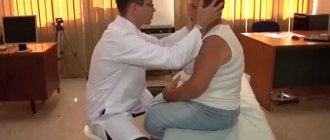
One of the most effective home exercises is the Epley maneuver. It is recommended for moving ear stones - otoliths - to a less sensitive area. Exercises are performed at home before bed for a week.
For the left ear (see picture): perform all exercises on the back for 30 seconds, then sit down for one minute. This cycle takes 2.5 minutes. For the right ear, the exercises are performed in a mirror manner. You need to do three cycles for each side.
Brandt-Daroff exercise
From the starting position while sitting, move left and right with your head slightly tilted. Stay in each position for 20-30 seconds. Perform the exercise several times a day.
***
Tips for those suffering from dizziness
● Avoid sharp bends and turns of the head. When you wake up, lie down for a while. Then slowly get out of bed. Sleep on a low pillow.
● Heavy physical activity is not for you.
● Avoid eating spicy foods.
● Get outdoors more often. Walking is good for you.
● It is dangerous to drive a car or work on moving machinery.
● Attractions with flashing lights and strong sound effects can cause an attack.
● Avoid stuffy rooms and direct sunlight.
● Consumption of alcohol and tobacco is unacceptable.
● Avoid stress, conflicts, psycho-emotional explosions. Try to establish friendly relationships with others.
Damage to the posterior semicircular tubule
Epley maneuver
The most studied is the Epley maneuver. It is used for pathologies of posterior and lateral SCC:
- The patient sits straight along the couch, with his head turned 45˚ towards the labyrinth that is being examined.
- The patient is placed in a lying position, while the head rotation is maintained, the head is tilted back slightly, hanging over the edge of the couch.
- After 20 seconds, the head turns to the healthy side by 90˚
- After 20 seconds, the head is turned in the same direction by 90˚ along with the patient's body, so that the face is facing down.
- After 20 seconds, the patient returns to a sitting position.
- The Simon maneuver is also used to treat posterior RCC lesions:
- In a sitting position, turn the head 45˚ towards the “healthy” ear, for example the right one
- The patient is quickly placed on his left side (head face up), an attack of dizziness occurs with rotatory nystagmus to the left, the position is maintained for 3 minutes. During this time, the otoliths descend to the lowest part of the RCC.
- Quickly turn the patient onto his right side (head face down). Maintain the position for 3 minutes.
- The patient is slowly returned to the starting position.
The fixed otolith resolves within a few weeks. The same amount of time is required for attacks of dizziness to disappear during the natural course of the disease.
According to research by Casani A.R. et al. (2011) the average duration of dizziness with damage to the posterior ACC was 39 days, with damage to the lateral ACC - 16 days.
Manipulations are often accompanied by a sharp temporary increase in the symptoms of the disease: dizziness, nausea, vegetative symptoms.
After the maneuver, the patient must be monitored after 3 days and 1 month, which will allow the maneuver to be repeated if it is ineffective or to promptly begin a search for other causes of dizziness if new symptoms appear.
Relapses occur relatively rarely (3.8 - 29% of cases).
Brandt-Daroff gymnastics
If the maneuvers performed by the doctor are ineffective, patients with damage to the posterior RCC are recommended to perform Brandt-Daroff gymnastics on their own:
- In the morning, after sleep, sit on the bed with your back straight (Position 1)
- Then you need to lie on your left (right) side with your head turned up at 45° (to maintain the correct angle, it is convenient to imagine a person standing next to you at a distance of 1.5 meters and keep your gaze on his face) (Position 2)
- Hold this position for 30 seconds or until the dizziness disappears
- Return to the starting position sitting on the bed
- Then you need to lie on the other side with your head turned up 45° (Position 2)
- Stay in this position for 30 seconds
- Return to the starting position sitting on the bed (Position 1)
Repeat the described exercise 5 times.
If dizziness does not occur during the exercise, it is advisable to perform it only the next morning. If dizziness occurs at least once in any position, then you need to perform the exercises at least two more times: in the afternoon and in the evening.
Benign positional vertigo
Benign positional vertigo
- one of the most common forms of dizziness. Despite this, it is often undiagnosed. Positional vertigo was first described by Robert Barany in 1921. In 1952, Dix and Hallpike suggested that this disease was associated with damage to the statolith organs, since the attack of vertigo is caused by turning the head, and developed a provocative diagnostic test.
Further pathomorphological studies showed that a basophilic substance is deposited on the dome of the semicircular canal in patients suffering from benign positional vertigo. It is assumed that in most cases the deposit appears from the statoconia of the degenerating statolithic membrane of the elliptical sac, which, under the influence of gravity in a critical position of the head, causes movement of the dome of the semicircular canal, which is the cause of vertigo.
Exercises for lesions of the lateral ACC
Vannucchi extended position method
Lying on your side with the affected ear facing up for 12 hours,
Barbecue method
The “barbecue” method - turning the patient 360˚ - the patient in a lying position is successively turned towards the healthy ear by 90˚ until he takes the starting position.
Lampert and Tiel-Wilck method (in the video above - a maneuver for damage to the right ear)
The patient's head is rotated 270˚ from the affected ear to the healthy one.
Special studies have shown the sufficient effectiveness of exercises performed by patients. There were no differences in the effectiveness of the maneuver in specialized clinics and in primary care settings.
Prevention methods
Headache and the diseases it signals can occur even in a previously healthy person. However, if you follow simple recommendations from doctors and pay attention to prevention, they can be avoided. At home, you can take the following measures to prevent headaches:
- monitor your posture both when walking and while working in a sitting position;
- give up bad habits, smoking and drinking alcohol;
- devote time to physical education every day, perform simple exercises in the fresh air;
- eat right, try to get enough vitamins.
The Clinical Brain Institute offers individual programs for the diagnosis and treatment of diseases that manifest themselves as pain in the neck and back of the head. It is important not to tolerate discomfort, as it may indicate the beginning of dangerous processes in the body. If you pay attention to the first symptoms and receive competent medical care in a timely manner, you can prevent further development of diseases and dangerous consequences.
Clinical Brain Institute Rating: 4/5 — 12 votes
Share article on social networks
Drug treatment
There are no drugs that have a direct effect on canal/cupolothiasis.
Drug treatment is advisable only for frequent attacks or during maneuvers.
Drugs are used that reduce the excitability of the vestibular system, both selectively and through a general sedative effect. The first include drugs with a vestibulolytic effect - blockers of H1 and H3 histamine receptors, cinnarizine, atarax, first generation antihistamines - diphenhydramine, pipolfen.
Evidence has been obtained in favor of reducing the intensity of dizziness when performing the Epley maneuver simultaneously with taking betahistine 24 mg x 2 times a day for a week.
Sedatives, most commonly benzodiazepine tranquilizers (diazepam), are used in hospital settings for the symptomatic treatment of severe recurrent attacks.
Diagnosis of dizziness
Diagnosis of dizziness
is relatively simple and is based primarily on the patient’s characteristic complaints. To confirm the diagnosis, the Hallpike positional test is used. Treatment of benign positional vertigo involves a rehabilitation maneuver. In this case, repeated movements are carried out in the plane of the affected semicircular canal. As a result, otolith deposits break off and fall into the vestibule of the labyrinth. The disappearance of these deposits from the canal leads to the cessation of dizziness. Drug treatment is ineffective, since available drugs (for example, meclozine) do not eliminate an acute attack of positional vertigo. In severe cases, surgical treatment is indicated.
Thus, benign positional vertigo
is one of the most common types of dizziness. The favorable prognosis and high effectiveness of treatment make timely diagnosis of this disease necessary.
Note!
- True vertigo
is the illusion of movement (usually rotation). - Isolated systemic vertigo
, not accompanied by focal neurological symptoms, in the vast majority of cases is the result of damage to the peripheral vestibular apparatus. - Benign positional vertigo
is one of the most common forms of vertigo. It can occur after a traumatic brain injury or as a result of a viral infection. - Treatment of benign positional vertigo
involves a rehabilitation maneuver.