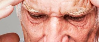
Dizziness: causes and treatment
(from the book Neurology. G.D. Weiss. Edited by M. Samuels. Translated from English - M., Praktika, 1997. -640 p.)
Dizziness is one of the most common and at the same time one of the most “disliked” complaints by doctors. The fact is that dizziness can be a symptom of a wide variety of neurological and mental diseases, diseases of the cardiovascular system, eyes and ears.
I. Definition. Since patients can call a variety of sensations “dizziness,” during the interview it is necessary first of all to clarify the nature of these sensations. They can usually be classified into one of four categories.
A. Vestibular dizziness (true dizziness, vertigo) is usually caused by damage to the peripheral or central part of the vestibular system. It manifests itself as the illusion of movement of one’s own body or surrounding objects. In this case, sensations of rotation, falling, tilting or swaying occur. Acute dizziness is often accompanied by autonomic symptoms (nausea, vomiting, increased sweating), anxiety, imbalance and nystagmus (the latter sometimes leading to blurred vision).
B. Fainting and pre-syncope. These terms refer to temporary loss of consciousness or the feeling of impending loss of consciousness. In a pre-fainting state, increased sweating, nausea, a feeling of fear and darkening of the eyes are often observed. The immediate cause of fainting is a drop in cerebral blood flow below the level necessary to supply the brain with glucose and oxygen. Fainting and presyncope usually develop against the background of arterial hypotension, heart disease or due to autonomic reactions, and the tactics for these conditions are completely different than for vestibular vertigo.
B. Impaired balance is characterized by instability, a wobbly (“drunk”) gait, but not true dizziness. The cause of this condition is damage to various parts of the nervous system that provide spatial coordination. However, patients with cerebellar, visual, extrapyramidal and proprioceptive disorders often define the feeling of unsteadiness as “dizziness.”
D. Vague sensations , often described as dizziness, occur with emotional disorders, such as hyperventilation syndrome, hypochondriacal or hysterical neurosis, depression. Patients usually complain of “brain fog,” feeling slightly intoxicated, lightheaded, or fear of falling. These sensations are quite clearly different from the sensations associated with vestibular dizziness, fainting and balance disorders. Since any dizziness, regardless of its cause, can cause anxiety, it cannot serve as evidence of the psychogenic nature of the disease.
D. Some patients with complaints of dizziness find it difficult to describe their sensations. In this case, it is advisable to conduct provocative tests.
1. The standard set of provocative tests for dizziness includes:
A. Orthostatic test. b. Forced hyperventilation for 3 minutes. V. Sharp turns while walking or spinning in a circle while standing. d. Nilen-Barany test for positional vertigo. d. Valsalva maneuver, which increases dizziness caused by craniovertebral anomalies (for example, Arnold-Chiari syndrome) or perilymphatic fistula, and also causes lightheadedness in patients with cardiovascular diseases.
2. After each test, it is necessary to ask whether the resulting dizziness resembles the sensation that worries the patient. For orthostatic hypotension, hyperventilation syndrome, positional vertigo and many vestibular disorders, test results are well reproducible, which provides important diagnostic information.
II. Clinical examination of patients with vestibular vertigo. In order to evaluate the results of research, it is necessary to have a good knowledge of the relationships of the vestibular system with the oculomotor, auditory and spinocerebellar systems. There are two main types of vestibular reflexes. Thanks to the vestibulo-ocular reflexes, gaze fixation on the objects under consideration is maintained, that is, the constancy of the image on the retina. Vestibulospinal reflexes provide the positioning of the head and torso necessary for coordinated movements and maintaining an upright posture.
A. Nystagmus in patients with dizziness is the most important sign of vestibular disorders. Knowing a few simple physiological principles can help you avoid common mistakes in interpreting nystagmus.
1. Canal-ocular reflexes. Each horizontal semicircular canal is connected through the neurons of the brain stem with the oculomotor muscles in such a way that a decrease in impulses from it causes the eyes to deviate towards this canal, and an increase causes movement in the opposite direction. Normally, the impulses constantly flowing into the brain stem from the right and left semicircular canals and otolith organs are equal in intensity. A sudden imbalance of vestibular afferentation causes a slow eye deviation that is interrupted by rapid cortical activation-induced corrective eye movements in the opposite direction (nystagmus).
2. Lesions of the labyrinth usually cause a decrease in impulses from one or more semicircular canals. In this regard, with acute unilateral lesions of the labyrinth, unidirectional nystagmus occurs, the slow phase of which is directed towards the affected ear, and the fast phase - in the opposite direction. Nystagmus can be rotatory or horizontal. It intensifies when the eyes are moved towards its fast phase (that is, towards the healthy ear). With acute vestibular dysfunction, surrounding objects usually “rotate” in the direction of the fast phase of nystagmus, and the body in the direction of the slow phase. Patients sometimes better determine the direction of rotation with their eyes closed. In a standing position, patients deviate and fall predominantly towards the slow phase of nystagmus (that is, the affected ear).
3. Central nystagmus. Alternating nystagmus, which changes its direction depending on the direction of gaze, is more often observed with drug intoxication, lesions of the brain stem, or pathological processes in the posterior cranial fossa. Vertical nystagmus almost always indicates damage to the brainstem or midline cerebellar structures.
B. Cold test. Normal physiological stimuli simultaneously affect both labyrinths. The value of the cold test is that it allows you to study the function of each labyrinth separately. The study is carried out with the patient lying down; the head is raised at an angle of 30°. The external auditory canal is washed with cold water, thereby simulating unilateral vestibular hypofunction (observed, for example, with vestibular neuronitis or labyrinthitis). Cold water causes movement of the endolymph, as a result of which the impulse from the horizontal semicircular canal decreases. Normally, this leads to nausea, dizziness and horizontal nystagmus, the slow phase of which is directed in the direction being examined, and the fast phase in the opposite direction. Monitor the direction, duration and amplitude of nystagmus. A decrease in response on one side indicates damage to the labyrinth, vestibulocochlear nerve, or vestibular nuclei on that side. The study is contraindicated if the eardrum is damaged.
B. Electronystagmography. The retina is negatively charged in relation to the cornea, so when the eyes move, the electric field changes and an electric current occurs. Recording this current (and therefore eye movements) using electrodes placed around the eyes is called electronystagmography. This method allows you to quantify the direction, speed and duration of nystagmus. Electronystagmography is used in functional vestibular tests to record spontaneous, positional, cold and rotational nystagmus. Electronystagmography can be used to record nystagmus with eyes closed. This provides important additional information since nystagmus is often suppressed during gaze fixation.
D. Hearing loss and tinnitus can occur with diseases of the peripheral vestibular system (inner ear or vestibulocochlear nerve), if the hearing aid is involved in the process. When the central nervous system is damaged, hearing is rarely impaired. For vestibular vertigo, audiological testing often helps establish the diagnosis.
1. In pure-tone audiometry, the threshold for the perception of sounds of different frequencies is measured. For differential diagnosis of sensorineural and conductive hearing loss, the auditory threshold for air and bone conduction of sound is compared.
2. For a more accurate audiological assessment, speech perception and intelligibility, the phenomenon of accelerated increase in sound volume and tone decay are additionally examined.
D. Stabilography - the study of balance using a moving platform - allows you to quantify involuntary postural reflexes that prevent falls, as well as the role of information from various senses in maintaining balance.
E. Functional vestibular tests, electronystagmography and stabilography are complex and labor-intensive procedures. They cannot replace a thorough clinical examination, and for non-vestibular vertigo they are not necessary.
III. Diagnosis and treatment of diseases accompanied by vestibular vertigo. The two most common causes of vestibular vertigo are vestibular neuronitis and benign positional vertigo.
A. Vestibular neuronitis (acute peripheral vestibulopathy, vestibular neuritis).
1. General information. Vestibular neuronitis is characterized by a sudden, prolonged attack of dizziness, often accompanied by nausea, vomiting, loss of balance, and a feeling of fear. Symptoms worsen with head movements or changes in body position. Patients tolerate this condition extremely hard and often do not get out of bed. Spontaneous nystagmus is characteristic, the slow phase of which is directed towards the affected ear. On the same side, the reaction to a cold test decreases. Positional nystagmus is often noted. Sometimes there is noise and a feeling of fullness in the ear. Hearing does not decrease, and the results of audiological examination remain normal. There are no focal symptoms indicating damage to the brain stem (paresis, diplopia, dysarthria, sensory disturbances). The disease occurs in adults of any age. Acute dizziness usually resolves spontaneously within a few hours, but may recur over the next few days or weeks. Subsequently, residual vestibular dysfunction may persist, manifested by imbalance, especially pronounced when walking. In almost half of cases, attacks of dizziness recur after several months or years. The cause of vestibular neuronitis is unknown. A viral etiology has been suggested (as with Bell's palsy), but there is no evidence of this. Vestibular neuronitis is more of a syndrome than a separate nosological form. Neurological and otoneurological examinations help establish the peripheral nature of vestibular dysfunction and exclude lesions of the central nervous system, which usually have a less favorable prognosis.
2. Therapeutic measures. A few simple techniques can significantly reduce dizziness.
1) Since head movements and external stimuli increase dizziness, the patient is recommended to lie in a darkened room for 1-2 days.
2) Gaze fixation reduces nystagmus and dizziness in peripheral vestibular disorders. Often the condition improves - and even to a greater extent than when lying with eyes closed - if patients fix their gaze on some nearby object (for example, on a picture or a raised finger).
3) Since mental stress increases dizziness, it is advisable to combine gaze fixation with mental relaxation methods.
4) In case of persistent vomiting, intravenous fluid administration is indicated to prevent dehydration.
5) Measures for persistent dizziness. With vestibular neuronitis, the condition does not improve significantly in the first 1-2 days. The person feels seriously ill and is afraid of repeated attacks of dizziness. In such a situation, it is important to reassure the patient, convincing him that vestibular neuronitis and most other acute vestibular disorders are not dangerous and pass quickly. It should also be explained that within a few days the nervous system will adapt to the imbalance between both vestibular organs (even if one of them is irreversibly damaged) and the dizziness will stop.
6) Vestibular gymnastics, which stimulates central compensatory processes, begins a few days after the acute manifestations subside.
B. Benign positional vertigo
1. General information. Benign positional vertigo is probably the most common vestibular disorder. Dizziness in this case appears only when moving or changing the position of the head, especially when tilting it back and forth. This condition often occurs when the patient turns over from his back to his side and suddenly, at a certain position of the head, feels that “the room has moved.” Dizziness usually lasts a few seconds. Often patients know in what position of the head it occurs. Changes in head position can worsen vertigo in vestibular neuronitis and many other peripheral or central vestibular disorders, but in benign positional vertigo, symptoms occur only with certain movements and are absent at other times.
2. Differences from positional vertigo of central origin. Positional vertigo can also occur with many other diseases, including lesions of the brain stem (multiple sclerosis, stroke or tumor). In order to distinguish benign positional vertigo from more dangerous diseases of the central nervous system, the Nilen-Barany test is performed. The seated patient is tilted with his head at an angle of 45°, after which he is lowered onto his back. Then the test is repeated, after first turning the thrown back head first to the right, then to the left. The result is assessed by the appearance of nystagmus and dizziness. The latent period, duration, direction and exhaustibility of nystagmus are of important diagnostic importance. In benign positional vertigo, the latent period of nystagmus and dizziness is several seconds, the nystagmus is rotatory, and its fast phase is usually directed towards the affected ear. Nystagmus and dizziness are usually short-lived (less than 30 s) and decrease with repetition of the test (depletion of nystagmus). The Nilen-Barany test can confirm the diagnosis of benign positional vertigo. However, a negative result does not exclude this disease, since its symptoms are transient and are not always triggered by head movement.
3. Etiology. Benign positional vertigo can occur after traumatic brain injury, a viral disease, otitis media or stapedectomy, as well as with certain intoxications (for example, alcohol and barbiturates). Idiopathic cases of the disease, apparently, in most cases are associated with cupulolithiasis, a degenerative process with the formation of otoconial deposits in the cupula of the frontal semicircular canal, resulting in a sharp increase in the sensitivity of this canal to gravitational influences when the position of the head changes.
4. The course of the disease can be very different. In many cases, symptoms resolve on their own within a few weeks and then do not return until months or years later. Sometimes a short-term attack occurs only once in a lifetime. Only rarely does positional vertigo persist for a long time.
5. Treatment. For symptomatic therapy, the above remedies are used, but they are often ineffective. With careful repetition of movements that provoke dizziness, pathological reactions are gradually “exhausted.” Some believe that vestibular exercises, which include provocative head movements, speed up recovery. Patients are advised to hold their head in a position that usually causes dizziness for 30 seconds. This simple exercise, performed 5 times every few hours, in most cases brings improvement within a few weeks. If such vestibular gymnastics is accompanied by too unpleasant sensations, then a soft corset is used that immobilizes the neck and prevents the head from tilting in an unfavorable direction. As with vestibular neuronitis, it is important to convince the patient that, despite the extremely unpleasant sensations, the disease will soon pass and is not life-threatening. It is extremely rare for severe persistent positional vertigo to transect the ampullary nerve coming from the frontal semicircular canal on the affected side.
B. Post-traumatic dizziness. Despite the fact that the labyrinth is protected by a bone sheath, its thin membranes are easily damaged by injury. Uncomplicated concussion is accompanied by dizziness in more than 20% of cases. With traumatic brain injury, transient autonomic disorders (palpitations, hot flashes, increased sweating), which are accompanied by non-vestibular dizziness, are also possible. Post-traumatic dizziness manifests itself in two main syndromes.
1. Acute post-traumatic dizziness. Vestibular dizziness, nausea and vomiting can occur immediately after injury due to the sudden shutdown of one of the labyrinths (labyrinth shock). Less commonly, dizziness is caused by transverse or longitudinal fractures of the temporal bone, which are accompanied, respectively, by hemorrhage in the middle ear or damage to the eardrum with bleeding from the external auditory canal.
Clinical picture. Dizziness is constant. Characterized by spontaneous nystagmus with a slow phase directed towards the lesion, and imbalance with a tendency to fall in the same direction. Symptoms intensify with sudden movements of the head and in a position where the damaged labyrinth is at the bottom.
2. Post-traumatic positional vertigo. Within a few days or weeks after the injury, repeated short-term attacks of vestibular dizziness and nausea may occur, which are provoked by head movement.
A. The clinical picture is the same as for benign positional vertigo.
b. Forecast. In most cases, spontaneous remission occurs within 2 months after injury, and within 2 years in almost all.
3. Perilymphatic fistula. The membranous labyrinth filled with endolymph is surrounded by the perilymphatic space. When a rupture occurs in the area of the oval or round opening, a perilymphatic fistula can form, through which changes in pressure in the middle ear cavity are directly transmitted to the inner ear. The cause of a perilymphatic fistula can be, in particular, barotrauma (from straining, sneezing, coughing, diving).
A. Clinical picture. Characterized by intermittent or positional vestibular vertigo and variable sensorineural hearing loss. Worsening often occurs with elevation (including rapid ascent in an elevator) and with physical exertion similar to the Valsalva maneuver (straining or lifting). Sometimes dizziness occurs with loud sounds (Tullio's symptom).
b. Diagnostics. A perilymphatic fistula should be suspected when vestibular or auditory disturbances appear after trauma. However, due to the variability of symptoms, it can be difficult to distinguish from other diseases (Meniere's syndrome, benign positional vertigo, craniovertebral anomalies). There are no pathognomonic signs in the study of pressor nystagmus, electronystagmography, or stabilography. Perilymphatic fistula is probably one of the common causes of vestibular vertigo of “unexplained etiology.”
V. Treatment. The perilymphatic fistula usually closes spontaneously, which is accompanied by the disappearance of symptoms. In persistent cases, if a perilymphatic fistula is suspected, surgical intervention is indicated (tympanotomy with restoration of the integrity of the oval or round foramen). After surgery, vestibular symptoms usually improve, but hearing is rarely restored.
G. Meniere's syndrome
1. General information. Meniere's syndrome usually begins between 20 and 40 years of age. It is characterized by sudden attacks of severe vestibular vertigo, lasting from a few minutes to several hours. Before an attack, and sometimes after it, there is a feeling of stuffiness and fullness, or noise in the ear, transient hearing loss. After an attack, imbalance may persist for a long time, especially noticeable when walking.
2. The course is characterized by remissions and exacerbations. At the onset of the disease, sensorineural hearing loss (mainly for low sounds) is episodic. As a result of repeated attacks, hearing progressively decreases, but periods of improvement are possible.
3. Pathogenesis. The main morphological changes in Meniere's syndrome are stretching of the walls and an increase in the volume of the endolymphatic space (endolymphatic hydrops). The cause may be impaired absorption of fluid in the endolymphatic sac or obstruction of the endolymphatic duct.
4. Treatment. During an attack, bed rest and vestibulolytic drugs are prescribed. The rational choice of drugs for the prevention of attacks and the assessment of their effectiveness are difficult due to insufficient knowledge about the pathogenesis of the disease and the unpredictability of its course (including the possibility of long-term spontaneous remissions). According to recent studies, any of the existing treatment regimens (including placebo) causes temporary improvement in approximately 70% of patients.
A low-sodium diet in combination with diuretics (thiazides or acetazolamide) has been recommended for the treatment of Meniere's syndrome; it has been suggested that this may reduce fluid accumulation in the endolymphatic space. However, the pathophysiological feasibility of this therapy has not been proven, and in recent years it has been used less frequently.
5. In a small proportion of cases, for frequent, severe, treatment-resistant attacks, surgical treatment is indicated. There is no ideal surgery for Meniere's syndrome. Shunting of the endolymphatic sac reduces dizziness in 70% of patients, but in 45%, hearing continues to decline after surgery. Destructive operations (selective transtemporal transection of the vestibular part of the vestibulocochlear nerve, labyrinthectomy or translabyrinthine vestibulectomy) are indicated for persistent severe dizziness and severe unilateral hearing loss.
6. Differential diagnosis
A. In all cases, it is necessary to exclude a tumor of the cerebellopontine angle (including schwannoma of the vestibular-cochlear nerve. Tumors of this location cause noise in the ear, hearing loss, imbalance, but only rarely - attacks of dizziness.
b. The cause of attacks of dizziness and hearing loss can also be infectious labyrinthitis, perilymphatic fistula, Cogan's syndrome, and hyperviscosity syndrome.
V. Congenital syphilis. Symptoms of labyrinthine lesions in congenital syphilis often appear only in middle age and can mimic Meniere's syndrome. Treponema pallidum persisting in the temporal bone causes chronic inflammation leading to endolymphatic hydrops and labyrinthine degeneration. The course is progressive. As a result, both ears are affected. All patients with bilateral Meniere-like symptoms should be examined for latent syphilis using treponemal reactions (primarily RIF-ABS), since non-treponemal reactions (including the reagin rapid test and the VDRL reaction) in syphilitic labyrinthitis can give negative results.
D. Labyrinthitis
1. Bacterial labyrinthitis. When there is a bacterial infection of the middle ear or mastoid process (such as chronic otitis media), bacterial toxins can cause inflammation of the structures of the inner ear (serous labyrinthitis). Symptoms may be minimal at first, but they gradually worsen without treatment. Direct infection of the labyrinth (purulent labyrinthitis) is possible with bacterial meningitis or disruption of the integrity of the membranes separating the inner ear from the middle ear. Patients experience acute vestibular dizziness, nausea, hearing loss, fever, headache and ear pain. Purulent labyrinthitis is a dangerous disease that requires early diagnosis and antibiotic therapy.
2. Viral labyrinthitis. Damage to the auditory and vestibular organs is observed with various viral infections, including influenza, herpes, rubella, mumps, viral hepatitis, measles, and infection caused by the Epstein-Barr virus. Most patients recover on their own.
E. Functional vertigo occurs as a result of a disruption in the interaction between the vestibular, visual and somatosensory systems, which normally work together to provide spatial orientation. Dizziness can also be caused by physiological stimulation of normally functioning sensory systems.
1. Motion sickness is caused by unusual acceleration of the body or a discrepancy between afferentation entering the brain from the vestibular and visual systems. In a person in a closed cabin on a ship or in the back seat of a moving car, vestibular afferentation creates a sensation of acceleration, while visual afferentation indicates the relative immobility of surrounding objects. The intensity of nausea and dizziness is directly proportional to the degree of sensory mismatch. Motion sickness is reduced when there is sufficient panoramic visibility to verify the reality of the movement.
2. Visually caused dizziness occurs when observing moving objects - due to a mismatch of visual afferentation with vestibular or somatosensory (for example, when a person watches a movie with a car chase).
3. High-altitude vertigo is a common phenomenon that occurs when the distance between a person and the stationary objects he observes exceeds a certain critical value. The often observed fear of heights prevents adaptation to the physiological mismatch of vestibular and visual afferentation.
G. Transient ischemia of the brainstem
1. General information
A. Clinical picture
1) Vestibular dizziness and imbalance are the two most common symptoms of transient brainstem ischemia resulting from damage to the arteries of the vertebrobasilar region. At the same time, only in rare cases are they the only manifestations of this disease. If repeated attacks of dizziness are not accompanied by other signs of brainstem ischemia (diplopia, dysarthria, sensory disturbances of the face or limbs, ataxia, hemiparesis, Horner's syndrome or hemianopsia), then they are usually caused not by vertebrobasilar insufficiency, but by peripheral vestibulopathy.
2) Impaired balance and blurred vision occur both with vestibular neuronitis and with lesions of the trunk, and therefore do not allow us to clarify the localization of the lesion. Acute hearing loss is not typical for ischemic lesions of the brainstem; a rare exception is occlusion of the anterior inferior cerebellar artery, from which the internal auditory artery arises to the inner ear.
b. Differential diagnosis
1) Since transient brainstem ischemia requires active therapy aimed at preventing brainstem stroke, it is important to differentiate it from more benign disorders (in particular, vestibular neuronitis).
2) In the interictal period with transient ischemia of the brainstem, there are no signs of focal brain damage. However, during an attack, careful examination can reveal disorders such as Horner's syndrome, slight strabismus, internuclear ophthalmoplegia, central alternating or vertical nystagmus, etc., characteristic of damage to the trunk, but not the vestibular apparatus. With ischemia of the trunk, it is often possible to induce positional nystagmus. The Nilen-Barany test helps to distinguish central from peripheral lesions. Vestibular vertigo and imbalance may also occur with brainstem lesions of other etiologies, such as multiple sclerosis or tumors.
H. Cerebellar stroke
1. Clinical picture. Damage to the cerebellum due to ischemia or hemorrhage in the posterior inferior cerebellar artery can manifest as severe vestibular vertigo and imbalance, which can easily be mistaken for symptoms of acute vestibular neuronitis. Sometimes the lesion is limited to the cerebellar hemisphere, and in this case there are no signs of damage to the lateral medulla oblongata (dysarthria, numbness and paresis of the facial muscles, Horner's syndrome, etc.). Infarction in the superior cerebellar artery causes abasia and ataxia, which are usually not accompanied by severe dizziness.
2. Diagnostics. Impaired balance with a tendency to fall towards the lesion is observed with damage to both the vestibular system and the cerebellar hemispheres and does not help in the differential diagnosis. Central alternating nystagmus, the fast phase of which is directed towards gaze, and hemiataxia indicate damage to the cerebellar hemisphere. A CT scan can diagnose cerebellar hemorrhage, but may not detect a heart attack (especially if the test is performed immediately after the onset of symptoms). A more reliable method for diagnosing cerebellar infarction is MRI.
3. Current. Cerebellar infarctions and hemorrhages are often limited in size and the outcome is favorable. Typically, gradual recovery occurs and the residual defect is minimal. More extensive lesions, accompanied by cerebellar edema, can cause compression of the trunk and fourth ventricle. This severe complication requires surgical decompression, but it can be prevented by timely dehydration, so early diagnosis and careful monitoring in the acute phase are extremely important for cerebellar strokes.
I. Oscillopsia is the illusion of vibration of stationary objects. Oscillopsia in combination with vertical nystagmus, instability and vestibular vertigo is observed with craniovertebral anomalies (for example, Arnold-Chiari syndrome) and degenerative lesions of the cerebellum (including olivopontocerebellar atrophy and multiple sclerosis).
K. Vestibular epilepsy. Dizziness can be the leading manifestation of simple and complex partial seizures if they occur in the vestibular areas of the cortex (superior temporal gyrus and association areas of the parietal lobe). Dizziness in this case is often accompanied by noise in the ear, nystagmus, and paresthesia in the contralateral limbs. The attacks are usually short-lived and can easily be confused with other diseases that manifest as vestibular vertigo. In most cases, such seizures are combined with typical manifestations of temporal lobe epilepsy. The diagnosis is confirmed by EEG changes. Treatment: anticonvulsants or resection of the affected area of the brain.
L. Migraine
1. Clinical picture. Dizziness can be a leading symptom of basilar migraine. During an attack, visual and sensory disturbances, disturbances of consciousness, and intense headache are also noted.
2. Diagnostics. Recurrent attacks of vestibular vertigo (in the absence of other symptoms) may be a manifestation of dissociated migraine. The diagnosis of migraine in this case is possible only if all other causes are excluded; it is more likely if there are other manifestations of this disease.
M. Chronic vestibular dysfunction
1. General information. The brain is able to correct the disrupted connection between vestibular, visual and proprioceptive signals. Thanks to central adaptation processes, acute dizziness, regardless of its cause, usually resolves within a few days. However, sometimes vestibular disorders are not compensated due to damage to the brain structures responsible for the vestibulo-ocular or vestibulospinal reflexes. In other cases, adaptation does not occur due to concomitant visual or proprioceptive impairments.
2. Treatment. Constant dizziness, impaired balance and coordination of movements can cause disability for the patient. Drug therapy in such cases is usually ineffective. Patients with persistent vestibular dysfunction are shown a set of special exercises (vestibular gymnastics).
A. Exercise goals
1) Reduce dizziness. 2) Improve your balance. 3) Restore self-confidence.
b. Standard complex of vestibular gymnastics
1) Exercises to develop vestibular adaptation are based on the repetition of certain movements or postures that cause dizziness or imbalance. It is believed that this should promote adaptation of the vestibular structures of the brain and inhibition of vestibular reactions.
2) Balance exercises are designed to improve coordination and use information from multiple senses to improve balance.
News
What is vertigo? This beautiful word denotes an unusual disease that is characterized by an unsteady, unsteady gait, falling, and dizziness in a person who feels like objects are spinning around him and feels like “the earth is spinning under his feet.” Attacks of dizziness are sometimes so intense that it is difficult for a person to stand still and maintain balance.
Vertigo can occur for a short time, due to external factors, for example, when motion sickness occurs while traveling in public transport or when visiting an amusement park or riding on a carousel.
Either be a symptom of diseases of the inner ear or problems with the center of balance in the brain, or be a symptom of a dangerous disease. For this reason, it is important to have information about this phenomenon in order to be fully armed to guard your own health.
Please note that if attacks of dizziness recur, be sure to consult a doctor!
Where does vertigo come from? With dizziness, there is a pronounced lack of coordination and balance. To find out its cause, one must understand how the transmission of nerve impulses occurs, which play a role in proprioception - the ability of a person to identify and sense the position of parts of his own body relative to each other and objects in the environment.
The inner ear is an organ that is responsible for a person's ability to maintain balance. It has a complex structure and consists of several sections: the vestibule, 3 semicircular canals and the cochlea. The inner ear is protected by bone tissue that forms the temporal lobe of the skull. It is filled with aqueous fluid - it moves when a person tilts and turns his head, and this information is perceived by sensitive cells to identify the position of the body in space.
The nervous system collects information that comes from the visual apparatus, tactile receptors and other sensory organs, as well as from the inner ear, and then analyzes it.
The center of balance is located in the cerebral cortex, namely in its temporal region. Along the nerves, impulses reach the vestibular nuclei - clusters of nerve cells capable of perceiving, analyzing and coordinating information received from different areas. Therefore, we know where is up and where is down, whether we are standing or moving, where our head is tilted, etc.
If at one of the stages the transfer of information is disrupted, a mismatch occurs - the person begins to feel dizzy. They imply any violation of the identification of body position in space, from mild disorientation to serious disorders of the vestibular apparatus.
Dizziness is not a disease, but a symptom of a huge number of conditions and pathologies. It occurs periodically in every person due to fatigue or hypovitaminosis, but its chronic manifestation, especially in combination with other symptoms (headache, nausea, vomiting, fainting) indicates the need for examination and treatment.
It is worth understanding that even minor disorders can indicate serious disorders of the nervous system in the initial stages, so self-medication is a dangerous and ineffective solution.
Main causes of vertigo.
Dizziness can be a symptom of a number of diseases and pathological conditions that lead to disruption of the vestibular apparatus. These are congenital or acquired disorders of varying severity, with different mechanisms of development and characteristics of clinical manifestations. Among the most common groups of causes of this symptom are the following:
- inflammatory diseases of the inner ear and vestibular nerve - can be of infectious or non-infectious origin;
- intoxication and poisoning , which lead to disruption of the vestibular apparatus;
- traumatic brain injuries - when certain areas of the cerebral cortex are damaged, dizziness may appear after a long time;
- metabolic disorders , including diabetes mellitus;
- psychogenic origin.
It is impossible to determine the cause of dizziness at home, especially if it is a consequence of organic lesions of the central nervous system.
Main symptoms of vertigo. Symptoms of dizziness may vary. In this regard, it is also customary to distinguish several varieties:
- tactile - the patient experiences a sensation of loss of support under his feet, instability, as on a ship;
- proprioceptive - associated with a change in the identification of the body in space, with a feeling that the body is rotating around an axis, and the surrounding objects remain in their places;
- visual - visually the human body remains motionless, and the environment begins to move in different projections.
Symptoms may last from a few minutes to several hours or more, and symptoms may recur at regular intervals or randomly.
Forms and types of vertigo.
To more accurately determine the cause of dizziness, there are several classifications of this symptom. This distribution is necessary to understand the danger of the disease and to prescribe an effective treatment regimen. Various types of therapy can be carried out by doctors of different specialties. So, the first classification distinguishes two types of dizziness:
- Systemic dizziness
- Unsystematic dizziness.
Next, we will examine each type of vertigo in more detail.
Systemic dizziness is a more dangerous type, which in any case indicates pathology. It is associated with a violation of nerve conduction at the level of the vestibular apparatus (inner ear, vestibular nerve, parts of the cerebral cortex). You can feel it when moving, both the person himself and objects relative to him. This condition may be accompanied by a feeling of panic, a general deterioration in the quality of hearing, and nystagmus.
The mechanism and causes of vertigo in this disease are compression and/or spasms of blood vessels that are involved in supplying the brain with oxygen. Due to oxygen starvation, dizziness occurs, often accompanied by headaches, which can ultimately lead to brain failure.
Systemic dizziness can be central, intermediate or peripheral, depending on the location of the pathological process.
Central vertigo (slow onset and not pronounced symptoms) has symptoms: rapid heartbeat; nausea; increased sweating.
Intermediate - the pathological process is localized at the stage of transmitting information from the inner ear to the central nervous system.
Peripheral vertigo (acute onset, sudden attacks that are difficult to tolerate) has symptoms: increased sweating; noise and ringing in the ears; nausea, often accompanied by vomiting; increased heart rate; hypotension. The most common cause of peripheral vertigo is a dysfunction of the inner ear, which can occur due to:
- high fluid pressure in the internal structure of the ear;
- toxic effects of certain medications, for example, antibiotics, as well as alcohol-containing drinks, tobacco products;
- improper functioning of blood vessels in the structure of the hearing organs;
- accumulation of calcium salts, it provokes aggravation of vertigo disease; in this case, dizziness appears only in certain positions of the neck and head, this condition can last only a few seconds;
- inflammatory processes of the hearing organs arising due to previous infectious or viral diseases
- ear injuries from sharp objects, accidental entry of small particles into the ear, as well as damage to the auditory nerve and tumor formations cause dizziness.
Systemic dizziness includes a number of diseases associated with various disorders of the conduction of nerve impulses to the cerebral cortex. The pathological process can be located at any level, and therefore the symptoms will differ. Thus, each pathology that may underlie the mechanism of development of dizziness should be considered separately. Systemic types occur in no more than a third of patients and require more detailed examination.
- Benign paroxysmal positional vertigo (BPPV) is the main cause of systemic varieties. The process is associated with the formation and deposition of crystals of calcium carbonate salts on the tissues of the semicircular canals of the inner ear. This is an acquired chronic disease, the leading clinical sign of which is short-term dizziness and loss of orientation in space. The attacks last no more than 1 minute, with no additional symptoms (headache, tinnitus).
- Inflammation of the vestibular nerve (vestibular neuronitis) is a dangerous condition that often develops against the background of a bacterial or viral infection. Its reproduction with damage to the nervous system causes a characteristic set of symptoms, which includes dizziness. The duration of their manifestation ranges from several hours to several days, and their intensity remains high or even intensifies. Hearing is preserved in vetibular neuritis.
- Post-traumatic dizziness is complete or partial disorientation that develops immediately after a traumatic brain injury. The process is associated with damage to areas of the cerebral cortex responsible for transmitting information about the position of the human body in space. Clinical signs may vary in intensity and persist until the tissue has completely healed, and in some cases they appear several days after the injury.
- Intoxication is another cause of dysfunction of the vestibular apparatus. Toxins, including aminoglycosides, can accumulate in peri- and endolymph, the fluid that fills the structures of the inner ear.
- Meniere's disease is a disease in which there is an increase in the amount of endolymph (hydrops). The fluid is located in the lumen of the labyrinth of the inner ear, and an increase in its volume leads to excess pressure on its walls. The disease occurs in attacks, the interval between which can range from several days to several months. The duration of one attack is from several hours to a day, and the symptoms are very pronounced. The patient experiences acute disorientation in space, dizziness and headaches. The disease progresses and causes gradual deterioration of hearing (usually a unilateral phenomenon), but complete loss does not occur.
- Temporal lobe epilepsy is a pathological condition that is manifested by a periodically manifested complex of autonomic disorders (dizziness, headache, low blood pressure, and others). The patient's condition deteriorates suddenly, for no apparent reason, and the attack may be accompanied by deterioration of vision.
- Lipotymic conditions are a set of symptoms that precede fainting. The patient experiences severe dizziness, weakness, his skin and mucous membranes turn pale, and his heartbeat becomes less frequent. An attack can occur for many reasons, including diseases of the cardiovascular system, a decrease in blood glucose concentration, an increase or decrease in blood pressure.
- Less commonly, dizziness can be associated with : head and neck injuries; problems with the brain (for example, stroke or cancer); taking medications that cause hearing impairment; migraines.
- Hemorrhage into the cerebellum also provokes vertigo. It can be diagnosed based on headaches, difficulty walking, and inability to move your gaze in the direction in which part of the cerebellum it occurred. In general, for a person with such a symptom, it is difficult to move his eyes from side to side.
- Multiple sclerosis. An age-related disease in which the patient cannot move his eyes strictly straight to the line of the nose and its tip.
- Complications caused by diabetes.
- Atherosclerosis (hardening of the arteries).
- Another cause of systemic vertigo is the toxic effects on the body of alcohol, drugs, tobacco products, as well as medications , for example, furosemide.
- Anemia and low hemoglobin levels.
Systemic dizziness most often occurs in old age, but can also occur in young people. The development of the disease should be monitored over time, taking into account the frequency and intensity of attacks. It is also important to monitor the occurrence of additional signs: hearing and vision impairment, fainting, nausea, migraines. These data will make it possible to determine the severity of violations even before specific analyzes are carried out.
Non-systemic dizziness is also called physiological. It manifests itself in the absence of pathological changes in the vestibular apparatus, but may be a consequence of its excessive irritation. Thus, non-systemic dizziness is one of the clinical signs of motion sickness syndrome, and also occurs after a long and monotonous rotation around its axis.
Non-systemic vertigo is characterized by darkening of the eyes, fainting, muscle weakness, poor health when blood pressure changes, and shortness of breath. One of the most characteristic signs of non-systemic dizziness is that its clinical manifestations intensify if you close your eyes. Additional clinical signs include general weakness and drowsiness, decreased visual acuity, deterioration of memory and attention. Similar symptoms may occur with systemic use of certain groups of drugs (benzodiazepines). The patient's condition in such cases improves by adjusting the dosage of medications.
A lack of oxygen in a stuffy and unventilated room can cause an attack of non-systemic vertigo; this phenomenon occurs very often.
Psychosomatics. Dizziness of psychogenic origin occurs with certain emotions and experiences - with fear, anxiety, neurosis. The main psychological causes of vertigo can be: moving, changing place of residence; loss of a loved one; unwillingness to accept and realize the realities of life here and now; new relationships; breakup of old relationships; job change.
It is worth understanding that systemic dizziness manifests itself in a more vivid clinical picture. It is associated with damage to neural connections, so symptoms appear and remain even without an apparent reason. In addition, loss of balance is accompanied by other characteristic manifestations of damage to the nervous system or analyzer systems. Non-systemic dizziness is temporary and can go away spontaneously as soon as the vestibular system adapts to environmental changes. The second case does not pose a danger to humans.
Why is vertigo dangerous? Due to dizziness, a person usually has a feeling of fear, although vertigo itself does not threaten life and health. The main thing is not to fall anywhere and not get hurt.
But for drivers, pilots, sailors, climbers or divers, vertigo can be fatal. It is especially dangerous for aircraft crews when the flight takes place over the sea, and in the aviators’ eyes water and sky merge, and top and bottom become confused. According to some reports, it was the pilot’s loss of orientation in space that led to the crash of the Tu-154 over the Black Sea in 2021.
What to do if you have an attack of vertigo?
- Move slowly. If you are standing or walking, lean on something stable. Breathe deeply. Don't rush anywhere - rest until your head becomes clearer.
- Don't look up or down. Looking up or down for long periods of time can increase disorientation and nausea. Keep your head and eyes straight and turn slowly.
- Do not look at moving objects, do not focus your gaze on very close or distant objects. This may make your dizziness worse.
- Lie in an inclined position. Try not to lie horizontally - this may worsen vertigo. Keep your head slightly elevated and sit or lie in an inclined position.
- Sit in silence. Avoid loud noises.
- Do the Epley maneuver or Foster maneuver. You can find out in detail about what these are and how to perform them on the Internet.
- See your doctor. If you once feel dizzy, most often there is no reason to worry. But if the feeling of vertigo recurs, be sure to consult a specialist.
Diagnosis of vertigo - vestibulo-atactic syndrome
Despite the fact that dizziness alone is not a dangerous symptom and cannot threaten the patient’s life, it can indicate serious problems with the vestibular system and organic lesions of the central nervous system in the early stages. It is impossible to cure these pathologies on your own, but an integrated and competent approach in a hospital setting will eliminate both the causes and manifestations of dizziness.
The diagnostic process is carried out by analyzing the functioning of the vestibular apparatus and the brain, after which the localization of the pathological process and its stage can be detected. During the initial examination, it is important to conduct a detailed interview with the patient and obtain a detailed description of the complaints. At this stage, it is possible to differentiate true dizziness from other conditions that are also often called by this term. Next, specialists will select an individual diagnostic scheme, which may include the following steps:
- examination of the spinal column (analysis of X-rays, CT or MRI) to identify osteochondrosis, vertebral displacements and other pathologies that cause impaired blood supply to parts of the brain;
- examination by a neurologist, which includes determination of nystagmus, various tests and assessment of the ability to coordinate movements;
- examination of the state of the brain using MRI or CT - carried out to exclude tumors and other diseases leading to demyelination of nerve fibers;
- electroencephalography is a way of assessing brain activity by capturing the signals it produces;
- study of the functioning of the vestibular apparatus - may include rotational tests, vestibulometry and other tests.
To make a diagnosis, neurologists also carry out: the Romberg test (the patient is placed with his arms and fingers extended forward, spread apart, then he is asked to close his eyes, if he begins to stagger and cannot maintain balance, this will tell the specialist about disturbances in the functioning of the cerebellum), the finger-nose test (touch finger to the nose with eyes closed, the diagnosis of vestibulo-atactic syndrome begins to be assumed with tremor of the fingers, inaccurate hit).
The intensity of the manifestations of the disease identified during the examination determines whether the diagnosis will be made - severe vestibulo-atactic syndrome, moderate or mild.
Treatment and prognosis.
When treating vertigo, it all depends on what caused the condition. In most cases of vertigo, treatment of vertigo is limited to conservative treatment, but there are cases when surgical intervention (for example, surgery on the inner ear) is necessary.
For isolated attacks of vertigo, medication is not always necessary, so you need to pay attention to some recommendations on how to get rid of dizziness at home:
Drink some water. Due to a lack of fluid in the body, you may feel dizzy because the blood volume decreases and the brain receives less oxygen.
Rest. Insufficient rest leads to general exhaustion of the body, which can subsequently cause an attack of vertigo.
Pills for motion sickness. This option should be used as a last resort, because drugs of this kind are addictive.
Ginger. This root was taken by sailors to combat seasickness. It helps improve blood circulation, which helps get rid of vertigo caused by oxygen starvation of the brain.
As with any other disease, in order for the treatment of vertigo to be most effective, it is first necessary to accurately determine the cause of this deviation. The main therapy prescribed for dizziness is taking medications that improve blood flow in the vessels of the brain, as well as improve blood flow to all parts of the brain. Similar drugs include Actovegin, Mildronate, Mexidol, Cavinton, Cerebrolysin, Trental, Vinpocetine, Tanakan, Vertigohel (homeopathy), Westinorm, Piracetam, Betaserc, Vertinex, Betagis, Fezam, etc.
In addition to these medications, various rehabilitation measures will improve the condition: exercises to develop balance, increase strength, coordination, stretching exercises for muscles and tendons.
Surgery. Allows you to get rid of vertigo radically - by removing the cause of the disease. Often used for brain tumors or middle ear tumors.
For example, an acoustic neuroma is a type of tumor of the nerve tissue of the ear that causes dizziness. However, surgery does not always solve the problem of such a symptom as dizziness. With injuries to the spine, neck and head, the head may feel dizzy either constantly or intermittently, and until the problem in the corresponding part of the neck or spine is resolved, attacks will occur again and again.
Treatment of vertigo with folk remedies:
It should be repeated again that it is difficult to determine the exact cause of vertigo, so at home, without knowing the main cause of dizziness, effective treatment cannot always be achieved immediately.
Folk remedies in the treatment of vertigo will most likely help in complex therapy. Typically, traditional healers recommend dietary supplements or plant-based medicines that improve blood flow and vascular function. Because with vertigo disease, it is especially important that blood flows well to all parts of the brain.
One such medicinal plant is Ginko Biloba . It improves the functioning of the vascular system of the brain and vestibular apparatus, due to this, blood flows well enough to the necessary organs, and the symptoms of vertigo disappear. In addition, preparations with Ginko Biloba extract have few contraindications.
Ginger is very effective for this disease . You can make an infusion with honey and ginger, it will perfectly relieve symptoms and calm the nervous system, and also prevent anxiety attacks. To prepare the infusion, you need to finely chop the ginger, one tbsp. l. grated ginger, pour boiling water over it and leave in a thermos for 6-8 hours. Take warm instead of tea with honey.
Freshly squeezed juices from beets and carrots are very useful for dizziness , which should be consumed before meals. Pomegranate juice also improves blood quality and promotes the production of red blood cells.
Drinking herbal tea with mint, clover, linden will calm the nervous system, resist attacks of vertigo and improve overall well-being. A tablespoon of dried mint (linden, clover) should be poured with boiling water and left to brew until it reaches a temperature suitable for drinking. It is better to drink this tea after meals.
Everyone knows such a plant as oregano , it helps with many diseases, vertigo disease is no exception. An infusion of oregano will ease your health in case of severe dizziness. To prepare the infusion, you need to take a handful of oregano inflorescences and stems and brew in a 500 ml thermos. and leave until the water cools down. Take half a glass.
Seaweed is rich in iodine and phosphorus essential for humans, which allow the human body to function properly and fully. If you regularly consume kelp, problems with the vestibular system can be avoided.
Garlic is very effective in treating vertigo - it reduces bad cholesterol in human blood, increases hemoglobin, and strengthens blood vessels. It is enough to consume 2 fresh cloves daily.
Please note that the effectiveness of treatment for vertigo and vestibulo-atactic syndrome, first of all, depends on the correct diagnosis and well-chosen methods. It is also very important to lead a healthy lifestyle, listen to your body and take care of yourself!
Be healthy and happy!
Control symptoms of acute vertigo with betahistine
Vertigo is always a symptom, not a disease, and it is a very common complaint in clinical practice, observed in 10–15% of patients who consult an otolaryngologist. The word “vertigo” refers to a variety of conditions: dizziness, imbalance, etc. At least 80 different diseases may include vertigo as a symptom [Oosterveld, 1985]. It is therefore not surprising that in approximately 40% of cases a definite and accurate diagnosis of vertigo is impossible, despite modern diagnostic procedures. Real vertigo is an unpleasant sensation of loss of balance, a feeling of spinning in space and disorientation. Nausea and vomiting usually accompany these sensations during an acute attack - it is no coincidence that many patients are afraid of attacks. Monitoring the condition of a patient suffering from vertigo is one of the “thorny” issues in medical practice. There are many ways to control vertigo; This includes pharmacotherapy, surgery, and rehabilitation of the vestibular apparatus. Pharmacotherapy. There are various medications available to treat vertigo. Betahistine is one of the most widely used anti-vertigo medications. It acts as a partial agonist at H1 receptors and as an antagonist at H3 receptors [Arrang, 1985; Venkataraman, 1998]. Various animal and human studies have shown that betahistine increases cochlear blood flow [Suga, 1969] as well as cerebral blood flow [Rivera, 1974]. The effectiveness of betahistine in the treatment of vertigo was compared with a placebo group; this has been demonstrated in various controlled clinical studies [Canty, 1981; Legend, 1988; Oosterveld, 1985]. The most interesting aspect of this drug is its dose-dependent effect, which has been established in many clinical studies. Unemoto et aI. studied the effects of betahistine in an electrophysiological study in cats and concluded that betahistine inhibits evoked electrical potentials in polysynaptic neurons of the lateral vestibular nucleus in a dose-dependent manner. Oosterveld (1987) studied the effect of different doses (8, 16, 32 mg) of betahistine on evoked vestibular nystagmus in a double-blind study on 10 healthy volunteers. The study showed that betahistine reduced the duration of evoked nystagmus in a dose-dependent manner. The higher the dose, the stronger the effect of betahistine. In our study, we assumed that betahistine has a dose-dependent effect. It has only been a few years since medicine abandoned the commonly prescribed dose of 8 mg 3 times a day. It was expected (given the dose-dependent effect of the drug) that a dosage of 16 mg 3 times a day would have a better effect. Based on this, our open-label, future-proof study of betahistine was undertaken to determine the efficacy and tolerance of betahistine as a vertigo medication at a dosage of 16 mg three times daily. Methods Patient selection Inclusion criteria The study included patients under 65 years of age who had experienced at least 1 episode of vertigo in the past month, including women who had adapted to any reliable form of contraception, as well as patients with hypertension and diabetes in a stable condition. Exclusion criteria The following patients were excluded from the study: patients with an infection of the inner or middle ear who required surgical intervention; those suffering from Parkinson's disease; with a brain tumor; with TBI; with epilepsy; with multiple sclerosis; with paralysis of the eye muscles; with psychological problems; pregnant and lactating women; with peptic ulcer; with bronchial asthma; with pheochromocytoma; patients being treated with systemic antihistamines; regular users of drugs or excessive amounts of alcohol. All patients who met the selection criteria were included in the study. Study Design This was an open-label, prospective, outpatient study of betahistine. A consent form was obtained from each patient prior to inclusion in the study. All patients received betahistine 16 mg 3 times daily after meals until remission or (maximum) 6 weeks. Any other medications used to treat vertigo were discontinued. Patients were included in the study one week after discontinuation of such drugs. Those with hypertension or diabetes who were stable continued to take their regular medications. Assessment At the "starting line" of the study, all patients underwent a clinical assessment, including a history of the underlying disease and any other disease from which the patient was currently suffering. It was noted how long the patient had suffered from attacks of vertigo in the past. The number of attacks in the last month was also recorded. Patients who had previously taken any other anti-vertigo drug were given a week of “respite” before the experiment. Demographic data was ascertained. Before the study, all patients underwent basic examination: complete blood count, audiogram, special audiological tests, radiological examination, if necessary. Follow-up assessments of patients were performed weekly throughout the betahistine treatment period. Three main parameters were assessed: frequency, duration and severity of vertigo attacks. The frequency of vertigo attacks was assessed as the number of attacks per week. The duration of vertigo attacks was recorded in seconds/minutes/hours. The severity of vertigo attacks was assessed on the following scale: • disabling (the person is bedridden); • severe (refusal from planned activities); • moderate (causes some interference in daily activities); • isolated (do not interfere with daily activities); • no seizures. Patients were classified according to the description of the symptoms of vertigo attacks as follows: • sudden, severe, short-term attack; • sudden, severe, gradually fading; • short attacks; • the condition is chronic, but not severe, persistent. The number of patients suffering from associated symptoms such as tinnitus, hearing loss, nausea, vomiting, fainting, and headache was noted. We also recorded other parameters - sugar (blood, urine), etc. Side effects (if observed) were recorded during each assessment session. Patients were asked whether they had drowsiness and whether the treatment was reducing their quality of life. Global Rating Patients and clinicians rated the overall effectiveness, tolerability, and impact of the drug on associated symptoms using a 4-point scale of excellent, good, fair, and poor. Results The study included 29 patients. Not a single patient dropped out of the experiment, and we analyzed the results for all 29 patients. Patient demographics are shown in Table 1. At baseline, the mean duration of illness (according to medical history) was 19.12 ± 38.25 weeks. Average number of attacks in the past month: 41.24±62.13. Average age in years: 42.86±14.44. Health status at the time of examination and concomitant diseases were recorded. Two patients had diabetes, four had hypertension, and three had spondylosis. The audiogram showed that 15 patients had bilateral hearing loss, 3 had unilateral hearing loss, and 11 patients had normal hearing. These patients did not have a single history of treatable vertigo. At the beginning of our experiment, 11 patients suffered from sudden, severe, short-term attacks of vertigo; 10 – from sudden, severe, gradually subsiding attacks of vertigo. Three patients had brief attacks; 5 had chronic, moderate in severity, persistent attacks of vertigo. At the baseline (starting) line of our study, the average frequency of vertigo attacks was 14.66±20.18 attacks per week; the average duration of the attack in minutes was 33.8±56.1. An average attack severity score of 2.9±0.62 points was recorded (Table 2). Frequency of vertigo attacks The average frequency of vertigo attacks at the baseline (starting) line of the study was 14.66 attacks per week; at the end of the first week of therapy, this number decreased statistically significantly (by 61.66%) to 5.62 attacks per week (p<0.05). After 3 weeks of therapy, the frequency of attacks decreased by 95.29% to 0.69 attacks per week, and this was highly statistically significant (p < 0.001) (Table 2, Fig. 1). Duration of vertigo attacks The average duration of vertigo attacks at the beginning of the study was 33.81 minutes. It decreased by 53.18% (p<0.05) – to 15.83 minutes. upon completion of the first week of therapy. After 3 weeks of therapy, the duration of attacks was reduced by 93.88% to 2.07 minutes, which was a significant difference (p < 0.001) (Table 2, Fig. 2). Severity of vertigo attacks At baseline, the mean attack severity score was 2.9. After the first week of treatment, the severity of attacks decreased by 36.9% - to 1.83 points. After three weeks of treatment, the attack severity score decreased by 95.17% – to 0.14 points (Table 2, Fig. 4). After completing 5 weeks of betahistine therapy, all of our patients were completely free of vertigo attacks. The average frequency, duration, and severity of attacks decreased dramatically. The effect of betahistine on associated symptoms is shown in Table 3 and Figure 3. 15 patients suffered from tinnitus. 14 patients got rid of this symptom after two weeks of treatment. After three weeks of therapy, none of the patients complained of tinnitus. Six patients complained of nausea/vomiting. Treatment with betahistine eliminated these associated symptoms in all patients within a week. Five patients complained of fainting before starting work. At the end of one week of therapy, syncope was not observed in 4 patients, and in the fifth, after two weeks of treatment with betahistine. At the baseline (starting) line, 20 patients complained of headaches. After treatment with betahistine, 18 patients no longer experienced headaches. We cannot assess the condition of the remaining two patients based on this symptom, since treatment was stopped after the vertigo resolved. At baseline, 13 patients complained of hearing loss. Three patients noted an improvement in their condition during betahistine therapy. We cannot report anything about improvement in hearing in the remaining 10 patients, since treatment was stopped after they were completely free of vertigo attacks. Table 4 shows the number of patients with vertigo attacks classified as “disabling”, “severe”, “moderate”, “isolated”; the assessment was based on weekly assessed severity of the condition. No adverse signs or symptoms were recorded in any patient. At the doses we used, no gastrointestinal side effects were noted, such as gastrointestinal upset, hyperacidosis, etc. No patient reported drowsiness or a negative impact on their quality of life. Overall assessment After completing therapy, 100% of patients rated the effectiveness of treatment, as well as tolerability of treatment, as “excellent” and “good.” 95% of patients considered the treatment effect on associated symptoms to be “excellent” or “good” (Table 5). After completion of betahistine therapy, clinicians rated the effectiveness and tolerability of treatment as either “excellent” or “good” for 100% of patients. Clinicians rated the effect of betahistine on associated symptoms as “excellent” or “good” in 95% of cases (Table 6). Subgroup analysis Patients with severe attacks of vertigo at baseline were also analyzed separately (n=18) (Table 7). The mean seizure frequency in this subgroup was 14.39. It was reduced over 2 weeks of treatment by 73% to 3.89. The average duration of vertigo attacks was 23.19 minutes. It was reduced by 99.4% to 0.15 min. within 2 weeks of therapy. The mean attack severity score was 3.0. It was reduced by 77.7% to 0.67 after 2 weeks of therapy. Clinically significant improvement was observed in this group of patients with severe attacks of vertigo, with the entire group becoming free of attacks after 3 weeks of treatment. Patients with disabling attacks of vertigo (n=4) were analyzed separately (Table 8). The average frequency of vertigo attacks in this group was 3.75 per week. It decreased after 2 weeks of treatment by 93.3% - to 0.25. The average duration of vertigo attacks was 82.75 minutes. It decreased by 63.8% to 30.0 minutes. after 2 weeks of therapy. The mean seizure severity score was 4.0. It decreased by 93.75% to 0.25 at the end of 2 weeks of treatment. All of these patients with disabling attacks of vertigo before treatment were completely free of attacks of vertigo after 3 weeks of treatment with betahistine. Discussion of results Various clinical studies have shown that betahistine is effective in relieving attacks of vertigo. It reduces the frequency of attacks, thereby providing a preventive effect. It also provides symptomatic relief by reducing the severity and duration of vertigo attacks. In the present study, betahistine was prescribed to patients at a dose of 16 mg 3 times a day. A statistically significant improvement was observed at the end of the 1st week of therapy. More than 93% reduction in the frequency, duration and severity of attacks was observed at the end of the 3rd week of betahistine therapy. All patients were completely free from vertigo attacks after completing 5 weeks of treatment with betahistine. Both clinicians and patients emphasized the effectiveness and good tolerability of betahistine in 100% of cases, giving ratings of “excellent” or “good.” The most interesting aspect of the study results is the analysis of the subgroup of patients with severe and disabling attacks (n=18+4) before the experiment. At the end of 2 weeks of therapy, 72% of patients with severe vertigo and 75% of patients with disabling vertigo were free of their symptoms. After 3 weeks of treatment with betahistine, all patients with severe and disabling vertigo were completely free of attacks. Another aspect that you have to think about when controlling attacks in a patient suffering from vertigo is the tolerability of drug therapy. In the present study, we observed that betahistine therapy at doses of 16 mg 3 times daily does not produce adverse symptoms or signs based on the experience of our patients. The medicine was specifically prescribed after meals. No patient discontinued treatment due to side effects. Both clinicians and patients considered the drug tolerability to be “excellent” or “good” (100% of patients). Thus, betahistine is very well tolerated in doses of 16 mg 3 times a day. Associated symptoms such as tinnitus, nausea and vomiting usually accompany attacks of vertigo. An interesting finding in our study was that all patients who complained of nausea and vomiting as associated symptoms were completely relieved of these symptoms after completing the first week of betahistine therapy. Other associated symptoms, such as tinnitus, fainting, and headaches, showed a statistically significant improvement during betahistine therapy. Most people with vertigo try to hold their head and neck rigidly and tensely, trying to prevent the attacks from getting worse. As a result, neck muscle spasm is one of the main causes of vertigo-related headaches. Relieving vertigo attacks leads to the release of this spasm and thus also successfully relieves headaches. As for the hearing loss, since in these cases it was chronic and not of the “fluctuating” type, it is not surprising that no noticeable improvement was noted in this symptom. Until recently, the medical trend was to control very severe attacks of vertigo or acute attacks with labyrinthine sedatives such as cinnarizine or prochlorperazine. Sedation or drowsiness is a common side effect of these drugs, which delay vestibular compensation [Kirtane, 1999]. But now that we have the experience of this study, we can take a new approach to managing such severe or acute attacks of vertigo. Betahistine 16 mg three times daily is the best choice for controlling acute or severe cases of vertigo. In the clinic of acute vertigo, which occurs largely due to imbalances in electrical potentials in the vestibular nuclei of both hemispheres, the following tendency is revealed: during the acute phase, cerebellar cells inhibit the vestibular nuclei and thereby alleviate symptoms. This phenomenon is very figuratively called “cerebellar clamp”. Gradually, over several weeks, the vestibular nuclei of the undamaged side take on the function of the affected nuclei. This is observed anatomically as the occurrence of cross-synapses and leads to a balance in the impulse activity of the nuclei of both sides. Once this happens, the vestibular nuclei are gradually released from the “cerebellar clamp” effect. This whole process is called "compensation"; thanks to it, a person or animal who has suffered from acute attacks of dizziness regains a sense of balance, even if the original pathology is irreversible. Most medications for dizziness cause sedation and CNS depression. This slows down the compensation process, which is so necessary for the patient's rehabilitation. And betahistine, which does not cause CNS depression, does not interfere with the compensation process. Betahistine, without being a sedative, does not disrupt the normal lifestyle of patients. It really has the effect of “acceleration” to vestibular adaptation, which is vital for the speedy rehabilitation of patients with acute peripheral vestibular disaster. Biswas (1998), Tighilet et al. (1995) made conclusions from the observations that the intake of betagistine per os (50 or 100 mg/kg of body weight) significantly accelerates recovery, restores statics and dynamics after interruption of the left -sided vestibular nerve in cats and thus significantly accelerates the vestibular compensation in cats. The conclusion in this way, the results of this study confirm the high efficiency of the treatment of betagistin at a dose of 16 mg 3 times a day with the control of patients with vertigo. Even acute or severe cases of vertigo can be successfully controlled with the help of such therapy. The results of the study also confirmed the excellent tolerance of therapy with betagistin, prescribed 48 mg per day (16 mg 3 times a day) to Indian patients; Such therapy did not cause sedations.
References 1. Arrang JM et aI., (1985) Actions of Betahistine at Histamine receptors in the brain. Euro. J. Pharmacol, 111, 73–84. 2. Biswas A. (1998) Drug Therapy in Vestibular Dysfunction In: 'An Introduction to Neurologist': 111. 3. Canty R et aI. (1981) Betahistine in peripheral vertigo. J. Laryngol OtoI, 95, 687–692. 4. Kirtane MV. (1999) Role of adaptation responses in clinical disorders. Ind J Otolarungol N NS, 51(2), 27–36. 5. Legent F et аI. (1988). Recurrent paraxosmal vertigo and sere: controlled clinical study. Concours Med, 29. 6. Oosterveld WJ (1985) Vertigo Current competitions in management. Drugs, 30, 275–283. 7. Oosterveld WJ (1987) Effect of Betahistine dihydrochloride on induced vestibular nуstagmus and double–blind studу. Clin Otolarungol, 12, 131–5. 8. Oosterveld WJ et al. (1989) Betahistine versus plastic in paraxosmal vertigo: a double–blind triаI. J Drug Ther Res, 14 (4), 122–126. 9. Rivera VM et aI. (1974) Vertebrobasilar arterial insufficiency with Dementia: ControIIed Trials of treatment with Betahistine Hydrochloride. J Am. Geriatr. Soc, 22, 397–406. 10. Suga F, Snow JB (1969). Coсhlear blood flow in response to vasodiIating drugs and some related agents. Larungoscore, 79, 1956–79. 11. Tighilet (1995). Betahistine ‚dihydrochloride treatment facilitates vestibular compensation in the cat. J Vest Res, 5(1), 53–66. 12. Unemoto N et aI. (1982) Inhibitory effect of Betahistine on PoIysunartis neurons in the Iatheral vestibular nucleus. Arch Otolarungol, 236, 229–236. 13. Venkataraman S (1998) Role of Betahistine in increasing cochlear and silver blood flow in vertigo patients. Neurosciences Toddy, 11(1, 2), 56–58.